
UW researchers uncover new clues about the cause of common birth defects
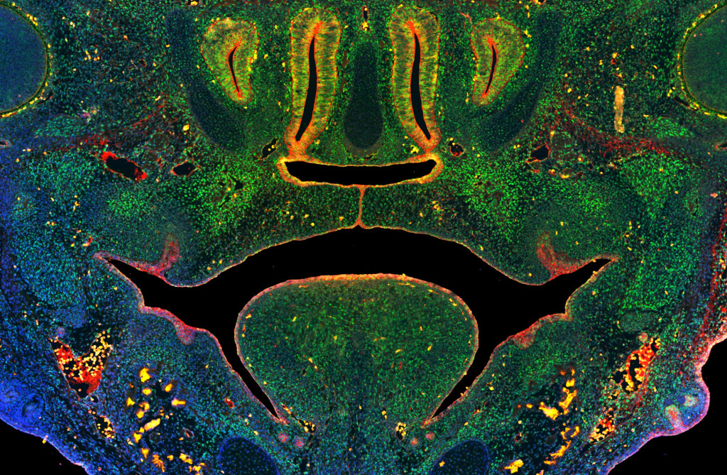
This image of a section through the midface of a mouse embryo illustrates fusion of the tissues that form the secondary palate above the tongue. Green staining illustrates cells expressing a key enzyme that mediates DNA methylation, blue indicates nuclei of all cells, red indicates epithelial cells.
Cleft lip and palate are the most common craniofacial birth defects in humans, affecting more than 175,000 newborns around the world each year. Yet despite decades of research, it’s still not known what causes most cases or what can be done to prevent them. But a recent study from the University of Wisconsin School of Veterinary Medicine (SVM) has uncovered new information about orofacial development in mice that researchers believe could one day help reduce the risk of these birth defects in humans.
Published this week in the Proceedings of the National Academy of Sciences (PNAS) the study provides the first direct evidence of a mechanism called DNA methylation being required for craniofacial development. DNA methylation is a process where a group of molecules are added to DNA that change the expression of genes without actually altering the DNA sequence. It’s also affected by various environmental factors. The researchers discovered that disruption to DNA methylation interferes with development of the lip and palate and causes these birth defects in mice.

Rob Lipinski
Led by Robert Lipinski, associate professor of comparative biosciences at the UW School of Veterinary Medicine, the research is an important step toward developing preventive strategies that could one day lessen the risk of cleft lip and palate, known collectively as orofacial clefts (OFCs), in both animals and humans.
“We knew from past research that genetics and the environment interact to cause these types of birth defects, but our understanding of the environmental component lagged far behind that of genetics.” says Lipinski. “Unlike genetics, we don’t have a permanent record of the prenatal environment that can be examined retrospectively, but connecting OFCs to DNA methylation helps narrow our focus on the particular environmental influences that modify the risk for these types of birth defects.”
His team’s work confirmed the essential role of DNA methylation in regulating orofacial development during embryonic development and demonstrates how disruptions to that process alter the ability of stem cells to form the connective tissue of craniofacial bone and cartilage, resulting in OFCs.
Lipinski and his team arrived at these results by first genetically manipulating DNA methylation in two separate groups of mouse embryos. The experiments resulted in seemingly contradictory results, with OFCs developing in one group of mice, but not the other. To understand why there was a difference between the groups, the team conducted another round of experiments in which they inhibited DNA methylation in mouse embryos at different stages of development. The timing of when DNA methylation occurs was critical to the development of orofacial clefts.
They found that exposure on the 10th gestational day resulted in OFCs but administering the same inhibitor just 48 hours later resulted in normal orofacial development.
Identifying this narrow window of gestational sensitivity is important, Lipinski says, because it not only helps narrow the focus of the next stage of his team’s research but it will also help design future public education initiatives once more is known about the modifiable environmental and behavioral risk factors that impact OFC risk in humans.
The 10th gestational day in mouse embryos corresponds with the beginning of the 5th week of embryonic development in humans–a stage at which many pregnancies may not yet be recognized.
“We know DNA methylation can be influenced by a variety of environmental factors, including maternal stress, diet, and exposure to drugs, toxins and environmental pollutants, and having a better understanding how orofacial development is regulated by environmentally sensitive mechanisms could directly inform birth defect prevention strategies,” he says. “This next phase of our team’s research is focused on identifying specific factors that influence DNA methylation during orofacial development and which could therefore alter OFC risk.”
Lipinski and his team are uniquely positioned to pursue this next stage of research because of another important outcome of the study: a new in vitro model the team developed. The model will allow them to rapidly screen thousands of dietary and environmental factors in a laboratory dish before testing the impact of specific factors on cleft susceptibility in mouse models.
The results in cell and animal models will help the researchers more quickly and accurately identify factors likely to be of consequence to human development.
Orofacial clefts of the upper lip and palate affect approximately 1 in 700 newborns, and individuals with OFCs navigate feeding difficulties as infants that require multiple surgeries, dental procedures, and speech therapy during childhood and adolescence. Studies have shown higher mortality rates at all stages of life for individuals with OFCs.
This study was supported by funding from the National Institutes of Health under award numbers R03DE027162, R56DE030917, RO1DE032710, U01 DK11807, and R01DK099328, and T32ES007015. Additional support was also provided by the University of Wisconsin Hilldale Undergraduate Research Award.
-
Interwoven
How Indigenous knowledge and science can work together to communicate about climate.
-
UW–Madison’s reach throughout Wisconsin adds up to $38.9 billion a year
The university supports more than 287,000 jobs and helps drive private-sector growth.
-
‘Being part of something bigger than you’ with Badger Volunteers
UW–Madison students head into the local community to find purpose and make a difference.
-
UW–Madison’s Tech Exploration Lab: Where the classroom meets the real world
The lab is built around a simple expectation: Students come to build, test and refine projects with real problems in mind.


