2025 Cool Science Image Contest
The 2025 Cool Science Image Contest is closed to submissions!
Winners were announced in October.
Want to share your work or interest in science? Get ready to share your coolest images and videos with us.
On this page:
What we’re looking for
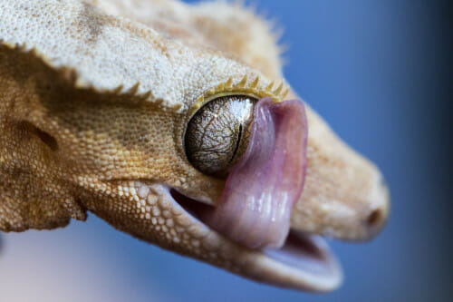
A crested gecko keeps its clear, immovable eyelids clean and moist with a swipe of the tongue in this 2020 winner by postdoc Nisha Iyer. Nisha Iyer
This contest is open to the UW–Madison community. Faculty, staff, students, postdocs and others with campus connections are eligible, and may enter as individuals or groups.
Images can depict an object or phenomenon from any discipline, and we welcome any method of producing an image — including, but not limited to:
- Microscopy
- Photography (astronomy, nature, etc.)
- Animations and (short) videos
- Medical imaging
- Science as art
- Schematics
- Photos of 3D-printed objects
Images will be judged on aesthetic and informational qualities. Check out the 2024 winners.
What you could win
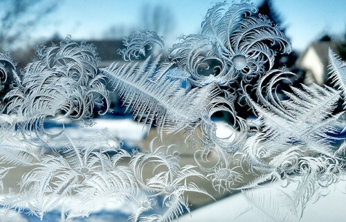
This cellphone photo of ice crystals on a windowpane by botany Professor Marisa Otegui was among the winner’s of the 2015 year’s Cool Science Image Contest. Marisa Otegui
- A published image. Winning entries are featured in slide shows on UW–Madison’s website and on select external web sites and venues.
- A large format print. Winners receive a large format print of their cool image or still from their video.
- Winning images will also be displayed in a fall semester exhibit at the McPherson Eye Research Institute’s Mandelbaum and Albert Vision Gallery and for a year at Promega’s headquarters.
How to enter
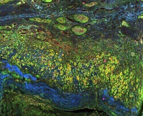
Immunohistology or impressionism? This image, by postdoc Wei-Hua Lee, of human tissue and blood vessels was among the winners of the 2016 Cool Science Image contest. Image by Wei-Hua Lee
The 2025 Cool Science Image Contest is closed to submissions.
The entry form will ask for the following information:
- Who to credit, including the names and titles of the individuals responsible for creating the image.
- Your affiliation with UW–Madison (i.e. undergraduate student, graduate student, postdoc, faculty, or staff). Please include university departments.
- Your permission to allow us to reprint your entries.
- A caption, no more than 200 words long, that answers questions like these:
- What does the image depict?
- How was the image made?
- What is an interesting fact about the object or phenomenon depicted?
- How is this object, phenomenon and/or method of image-making important to your research, discipline, studies or interests?
Please write your caption in layman’s terms, avoiding scientific jargon. It should be easy for a non-scientist to understand. An image that can be described in a meaningful way to a broad audience has an advantage in our contest.
Image quality is important. Please enter high-resolution image files. We are keenly aware of copyright issues on the web, and we always credit creators when publishing images or videos. We hope this encourages others to be respectful of copyrighted property.
Winners will be announced this summer.
Judges and advisors
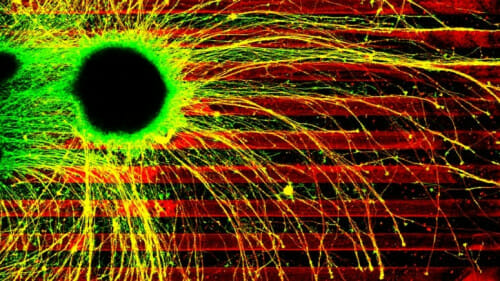
A 2021 contest winner shows the yellow connecting arms, called axons, of diseased human brain cells growing willy-nilly across boundaries of inhibitory chemicals (the red stripes). Healthy axons would precisely follow the dark lanes, giving researchers the opportunity to test the effects of disease-causing mutations on axon growth. Timothy Catlett graduate student, Cell and Molecular Biology; Timothy Gomez, professor, Neuroscience
- Steve Ackerman, professor of atmospheric and oceanic sciences
- Terry Devitt, emeritus director of research communications
- Kevin Eliceiri, director, Laboratory for Optical and Computational Instrumentation
- Michael King, visual communications specialist, College of Agricultural and Life Sciences
- Kara Rogers, science writer and editor, Encyclopedia Britannica
- Ahna Skop, professor of genetics
- Kelly Tyrrell, assistant vice chancellor, content strategy , Office of Strategic Communication
Tags: research
-
UW–Madison again named a ‘new Ivy’ by Forbes
The university was selected as one of the best in the country at "preparing and graduating the talent" that meets today's workforce needs.
-
UW–Madison graduate programs ranked among the nation’s best by U.S. News
Nearly 20 programs ranked in their respective Top 10 lists, shining a light on the breadth and depth of the university’s graduate offerings.
-
UW–Madison students ‘step into the professional world with a purpose’
More than 90% of UW graduates say their education prepared them for their career, according to new survey results.
-
How UW–Madison and industry are working together to shape Wisconsin’s AI future
As businesses explore how to apply AI effectively and responsibly, UW researchers, faculty and students are offering their knowledge in the emerging space.