
Science meets art: 2015 Cool Science Images unveiled
Whether a close-up of a leafcutter ant, or a micrograph of the neurons derived from marmoset stem cells, or an MRI of the hidden pathways in the human brain, submissions to UW–Madison’s 2015 Cool Science Image Contest continue to put science and nature on eye-catching display.
Sorting through a record number of entries, judges for the contest selected 11 still images and one video as winners of the annual competition. The judges — representing broad expertise in scientific imaging, art and science communication — worked through 115 submissions to arrive at this year’s winning entries.
Submissions overall and winning submissions represent a wide segment of the UW–Madison community, including faculty, staff and students and a range of disciplines from art to zoology.
“This year’s contributions were among the strongest we’ve had and reflect the diversity and creativity of the UW–Madison scientific imaging community,” says Kevin Eliceiri, a contest judge and director of UW–Madison’s Laboratory for Optical and Computational Instrumentation. “I was struck in particular by the broad number of disciplines and range of techniques represented.”
Winning images depended on techniques ranging from MRI to a cell phone camera.
2015 CSI Contest winners are:
Zoology graduate student Hilary Bultman for her micrograph of thyme plant floral trichromes.
Tyler Gordon, graduate art student, for his photograph, taken with the aid of a polariscope, of “Prince Rupert’s Drops,” a style of glass sculpture.
Caleb Weisnicht, undergraduate art student, and Andrew Klapper, undergraduate engineering student, for their compilation image of photographs of fungi found in Wisconsin forests.
Medical microbiology and immunology postdoctoral fellow Sabrina Koehler for her picture of a European beewolf and its honeybee prey.
Jackson Hetue of the Wisconsin Fast Plants Program in the Department of Plant Pathology, for his time-lapse video of germinating seedlings.
Alex Orellana, graduate student in art, and Brian Allen, graduate student in psychology, for their MRI of the human brain pathways involved in amblyopia or “lazy eye.”
Jorden Manasse, a postdoctoral fellow in the School of Veterinary Medicine’s Department of Pathobiological Sciences, for her micrograph of a stained cholelith or gall bladder stone in a golden lion tamarin.
Scott Vermilyea, of the Neuroscience Training Program in the School of Medicine and Public Health, neurobiology undergraduate Scott Guthrie, and Ted Golos and Marina Emborg, both professors in the School of Medicine and Public Health and researchers at the Wisconsin National Primate Research Center, for their micrograph of marmoset embryonic stem cells forming neurons.
Eshwar Udho, a postdoctoral fellow at the Waisman Center, for his micrograph of cultured mouse neurons from the lab of pediatrics Professor Pelin Cengiz.
Botany Professor Marisa Otegui for her cellphone picture of ice crystals on a windowpane.
Jessica Plavicki, an assistant scientist in the School of Pharmacy, for her micrograph of vasculature in the adult zebrafish brain.
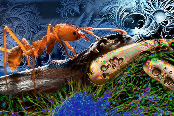
Volunteer photographer Don Parsons, for his portrait of a leafcutter ant in the lab of bacteriology Professor Cameron Currie.
The Cool Science Image Contest is sponsored by Promega Corp. Founded in 1978 in Madison, Promega is one of Wisconsin’s leading life sciences companies. Additional support is provided by DoIT’s Digital Publishing and Printing Services. The purpose of the contest is to provide a showcase for the work of Wisconsin researchers. All of the winning images and videos will be featured in an exhibit to be held during the fall semester at the Mandelbaum & Albert Vision Gallery, part of UW–Madison’s McPherson Eye Research Institute.
Tags: arts, contests, faculty awards, research, staff awards, student awards
-
Interwoven
How Indigenous knowledge and science can work together to communicate about climate.
-
UW–Madison’s reach throughout Wisconsin adds up to $38.9 billion a year
The university supports more than 287,000 jobs and helps drive private-sector growth.
-
‘Being part of something bigger than you’ with Badger Volunteers
UW–Madison students head into the local community to find purpose and make a difference.
-
UW–Madison’s Tech Exploration Lab: Where the classroom meets the real world
The lab is built around a simple expectation: Students come to build, test and refine projects with real problems in mind.


