Patient-derived induced stem cells retain disease traits
When neurons started dying in Clive Svendsen‘s lab dishes, he couldn’t have been more pleased.
The dying cells — the same type lost in patients with the devastating neurological disease spinal muscular atrophy — confirmed that the UW–Madison stem cell biologist had recreated the hallmarks of a genetic disorder in the lab, using stem cells derived from a patient. By allowing scientists the unparalleled opportunity to watch the course of a disease unfold in a lab dish, the work marks an enormous step forward in being able to study and develop new therapies for genetic diseases.
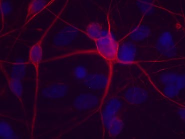
The nerves that control muscles, known as motor neurons (shown here in red), are lost in the devastating genetic disease called spinal muscular atrophy, causing weakness, paralysis, and early death. A team of UW–Madison stem cell biologists recreated the hallmarks of this disease in the lab using genetically reprogrammed stem cells created from a young SMA patient’s skin. The work gives scientists the opportunity to study the full progression of a disease in the lab and should improve understanding and treatment of genetic disorders. The motor neurons shown here were grown from cells from the patient’s healthy mother.
Photo: courtesy Clive Svendsen
As reported this week in the journal Nature, Svendsen and colleagues at UW–Madison and the University of Missouri-Columbia created disease-specific stem cells by genetically reprogramming skin cells from a patient with spinal muscular atrophy, or SMA. In this inherited disease, the most common genetic cause of infant mortality, a mutation leads to the death of the nerves that control skeletal muscles, causing muscle weakness, paralysis, and ultimately death, usually by age two.
Genetic reprogramming of skin cells, first reported in late 2007 by UW–Madison stem cell biologists James Thomson and Junying Yu and a Japanese group led by Shinya Yamanaka, turns back the cells’ developmental clock and returns them to an embryonic-like state from which they can become any of the body’s 220 different cell types. The resulting induced pluripotent stem cells, known as iPS cells, harness the blank-slate developmental potential of embryonic stem cells without the embryo and have been heralded as a powerful potential way to study development and disease.
Just one year later, the new work is fulfilling that promise.
“When scientists study diseases in humans, they can normally only look at the tissues affected after death and then try to work out — how did that disease happen? It’s a little like the police arriving at the scene of a road accident — the car’s in the ditch, but they don’t know how it got there or the cause of it,” explains Svendsen, a professor of anatomy and neurology in the UW–Madison School of Medicine and Public Health and the Waisman Center, and co-director of the Stem Cell and Regenerative Medicine Center.
With iPS cells, he says, “Now you can replay the human disease over and over in the dish and ask what are the very early steps that began the process. It’s an incredibly powerful new tool.”
In the new study, the researchers created iPS cells from stored skin cells of a young SMA patient and his mother, who does not have the disease. The cells grew well in the lab, and the group developed a new method to efficiently drive them to make large numbers of motor neurons, the cells that control muscles and that are affected in SMA.
Initially, the motor neurons thrived in both samples. But after about a month, “the accident started happening,” Svendsen says, and the motor neurons from the patient-derived cells began to disappear.
“The motor neurons we got started to die in culture, just like they do in the disease. This is the first validation of a human disease that we’ve modeled in a culture dish,” he says.
They can now begin to dissect what kills the motor neurons and why these cells alone are targeted in the disease. Past studies to understand the effects of the SMA-causing mutation have often relied on the easy-to-obtain skin cells, which are not affected in SMA and offer limited insight into how and why motor neurons die, says UW–Madison researcher Allison Ebert, lead author on the new study.
“If we start to understand more of the mechanism of why the motor neurons specifically affected in the disease are dying, then potentially new therapies can be developed to intervene at particular times early in development,” she explains. Current SMA treatment options are limited, and there is no cure.
Ebert points out that the patient-derived iPS cells can offer scientific advantages over other approaches, including embryonic stem cells, for studying disease. In effect, the researchers can watch the unfolding of an accident that has already occurred, and the known clinical outcome — the course and severity of the patient’s disease — should help them understand how the changes they see in the cells fit into the bigger picture of the disease.
“The development of human-derived SMA motor neurons is an important step forward for the SMA field, especially as a variety of therapeutic avenues are being examined,” agrees SMA expert Christian Lorson, a professor of veterinary pathobiology at MU and an author on the paper. “To be able to investigate therapeutic activity in these cells, whether it be novel drugs, viral vectors, oligonucleotides, or a better understanding of disease pathology, the iPS SMA motor neurons represent an excellent disease-related context.”
While complex and late-hitting disorders like Alzheimer’s and Parkinson’s diseases will be harder to model with iPS cells, the researchers say the approach should pave the way for studies of other genetic disorders, such as Huntington’s disease. “We have to find better ways to model complex human diseases that are difficult to reproduce in animals,” Svendsen says. “iPS cells represent a promising new research tool to reach this goal.”
He credits the UW–Madison Stem Cell and Regenerative Medicine Center with facilitating the work, especially by drawing on the expertise of Yu and Thomson, who pioneered the technique, to create the iPS cells used in this study. “This is an example of how the center is working to collaborate on campus and off campus to bring these kind of things to fruition,” he says.
# # #
- Jill Sakai, 608-262-9772, jasakai@wisc.edu
Tags: business, health & medicine, research, stem cells
-
UW–Madison’s reach throughout Wisconsin adds up to $38.9 billion a year
The university supports more than 287,000 jobs and helps drive private-sector growth.
-
‘Being part of something bigger than you’ with Badger Volunteers
UW–Madison students head into the local community to find purpose and make a difference.
-
UW–Madison again named a ‘new Ivy’ by Forbes
The university was selected as one of the best in the country at "preparing and graduating the talent" that meets today's workforce needs.
-
UW–Madison graduate programs ranked among the nation’s best by U.S. News
Nearly 20 programs ranked in their respective Top 10 lists, shining a light on the breadth and depth of the university’s graduate offerings.