University spinoff aims to hit the mark precisely with brain-scanning tool
As brain surgeons test new procedures and drugs to treat conditions ranging from psychiatric disorders to brain cancer, accuracy is becoming an ever-greater issue.
In treating the brain, the state of the art today starts with images from a magnetic resonance (MR) scanner, usually made a few days before surgery. Then, in the operating room, multiple cameras track instruments as they are inserted through a hole in the skull, creating images that can be superimposed on the original MR scans.

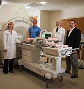
Walter Block
But there is no guarantee that the brain will not shift slightly during the surgery and throw off the best efforts at exact guidance.
For 20 years, neurosurgeons have discussed a radical way to achieve real-time accuracy in placement: performing surgery with the brain inside an MR machine, says Walter Block, professor of biomedical engineering at the University of Wisconsin–Madison. “When you open the brain for surgery, the tissue can shift slightly, and that will throw off predictions made in advance.”
To bring the full promise of MR into the operating room, Block has formed a company called InseRT MRI to develop software that allows surgeons to observe the brain in real time on an MR machine during surgery.
Such a system would have a number of applications, he says. Drugs for brain cancer can be delivered over as long as 54 hours. “It would be valuable to see where the drug is going during the first few hours,” Block says. “Drugs move at different rates through gray and white matter, and this ability to recalibrate the treatment plan, based on actual data on where the drug is moving, would allow you to alter the location of the catheter or the flow rate of the medication.”

Marvel Medtech in Cross Plains, Wisconsin, is developing a system that would employ InseRT MRI’s guidance to biopsy breast tumors.
Photo: Center for Technology Commercialization
To get that accuracy advantage, Block does not envision forcing surgeons to learn a new operating environment. “Surgeons have operating room tools and work stations that are familiar to them,” he says. “We are creating a set of tools that make the MR space a comfortable place for the surgeon.”
UW-Madison neurosurgeon Azam Ahmed plans to use the system through test procedures on animal brains and cadavers, Block says. “We are working with Dr. Ahmed to design the workflow so it’s intuitive to him. We are not going to piggyback on top of a large scanner market designed for largely diagnostic purposes, kludging it to make it work for interventional applications.”
The goal is not to develop software that could be spliced into MR manufacturers’ systems, he says, “since every time they alter their software, we would have to change ours.” Instead, Block is borrowing tactics from the smartphone industry. “People write apps that use various phone resources — GPS, the screen, the orientation system. We look at the MR scanner as a set of resources that we can control. An app writer does not have to go to Samsung or Apple and say, ‘We have this idea.'”
Block says his software will interact with the MR machine through a software “portal” being developed by another firm.
“When you open the brain for surgery, the tissue can shift slightly, and that will throw off predictions made in advance.”
Walter Block
One obvious market is the pharmaceutical industry. “Any drug trial in the brain will cost hundreds of millions of dollars,” he says, “and we often see trials being repeated after post-mortem analysis raises questions about the accuracy of drug placement.”
Targeted surgery could also help remove bits of brain tissue to treat severe epilepsy. Marvel Medtech in Cross Plains, Wisconsin, is developing a system that would employ InseRT MRI’s guidance to biopsy breast tumors. The technology also raises the potential for localized psychiatric drug therapy, Block says.
In the brain, the MR-guidance system is already accurate to less than a millimeter, Block says. While conventional stereotactic systems can approach that accuracy “in the best case,” the error can rise to 1.5 or 2 millimeters — a vast distance in an organ as delicate as the human brain, in which damage to healthy tissue must be minimized.
Block says InseRT MRI’s competitive advantage resides in his long experience in medical imaging. “Our value is (faster) time to market. We have come up with ways to circumvent the significant hurdles that now limit image-guided therapy, and we believe we can do this faster than anybody else.”
