
Photo gallery The 2022 winners: Cool Science Image Contest
Winners in the University of Wisconsin–Madison’s annual Cool Science Image Contest — including a quilt organized around a mathematical theorem, a painting of tiny swimming plankton out for their daily constitutional and lung X-rays rendered by artificial intelligence as classic works of art — are some of the most diverse representations of science in the contest’s 12-year history.
A panel of eight experienced artists, scientists and science communicators chose nine more images and a video based on the aesthetic, creative and scientific qualities distinguished from scores of entries. The winning entries showcase animal cells, crystalline structure, quantum computing equipment and a sweeping view of our galaxy.
An exhibit featuring the winners is open to the public at the McPherson Eye Research Institute’s Mandelbaum and Albert Family Vision Gallery on the ninth floor of the Wisconsin Institutes for Medical Research, 1111 Highland Ave., through December. A reception including contest entrants will be held at the gallery Thursday, Sept. 29, from 4:30 to 6:30 p.m. and is also open to the public.
Winning creators used a wide array of tools, including incredibly sophisticated microscopes and point-and-shoot digital cameras, innovative machine learning computers and a needle and fabric. A common thread runs through the creators’ desire to explore the world around us with more than the mind’s eye.
“The pursuit of science is about more than abstract ideas,” says Kelly Tyrrell, a contest judge, molecular biologist and UW–Madison director of media relations and strategic communications. “Science can allow us to see the unseeable and uncover the unknown. Science images, videos and art can offer a tangible glimpse into and through the universe, and help us to better know ourselves, too.”
Story continues after gallery
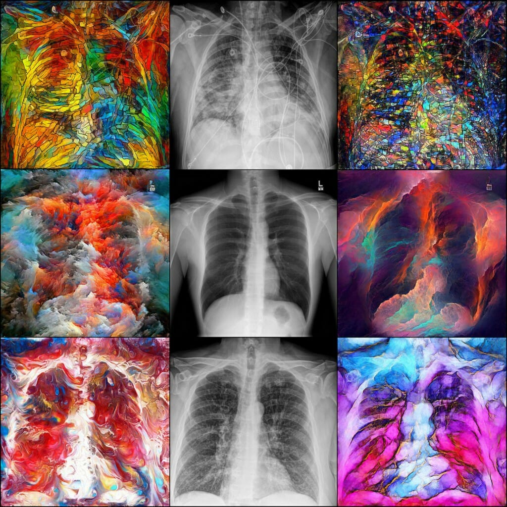
These stylistically different takes on chest X-rays were created by generative adversarial networks, computing networks designed to “learn” like the human brain learns. While these particular works of art are more beautiful than useful in radiology, GANs are used in medical imaging to enhance, classify and reconstruct information and understand the differences between X-rays of a COVID case (top), pneumonia (bottom) and healthy lungs (center).
Dalton Griner,
graduate student, Medical Physics;
Xin Tie,
graduate student, Medical Physics
chest x-rays and PyTorch deep learning framework
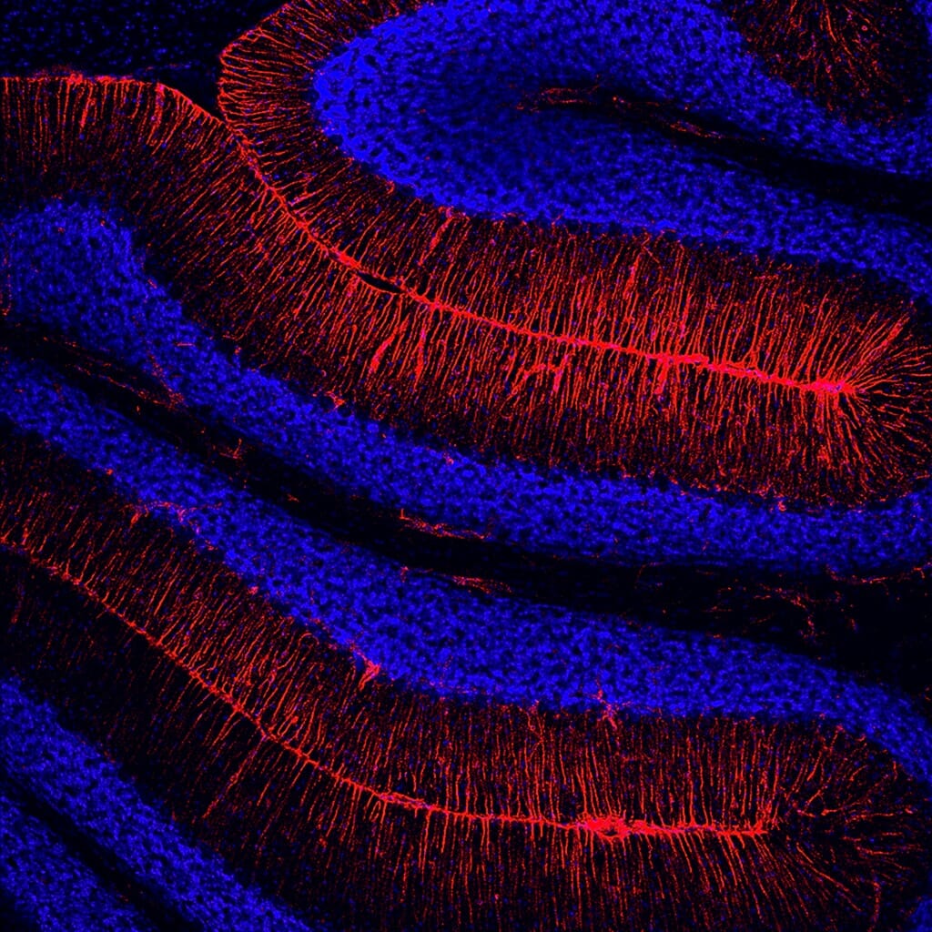
This image of brain cells from the cerebellum of a mouse was made possible by the addition of a gene that fuses a fluorescent red molecule to a protein called vimentin, which forms filaments in cell walls and is particularly present during cell development, wound healing and the spread of cancer. The red “tag” allows researchers to tell the blue nuclei from the red vimentin filaments in studies of the mice.
Karolina Lungova,
research intern, Neuroscience;
Darcie Moore,
professor, Neuroscience
confocal microscope
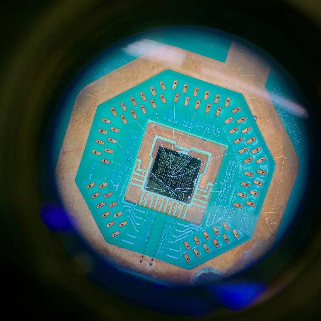
Viewed through the eyepiece of a microscope, aluminum wires one-third the diameter of a human hair connect superconducting devices to a microchip. During experiments, the microchip is cooled to -460 degrees Fahrenheit (just one-hundredth of a degree above absolute zero) to explore the laws of quantum physics and test nanotechnology for quantum computing.
Benjamin Harpt,
graduate student, Physics
digital camera
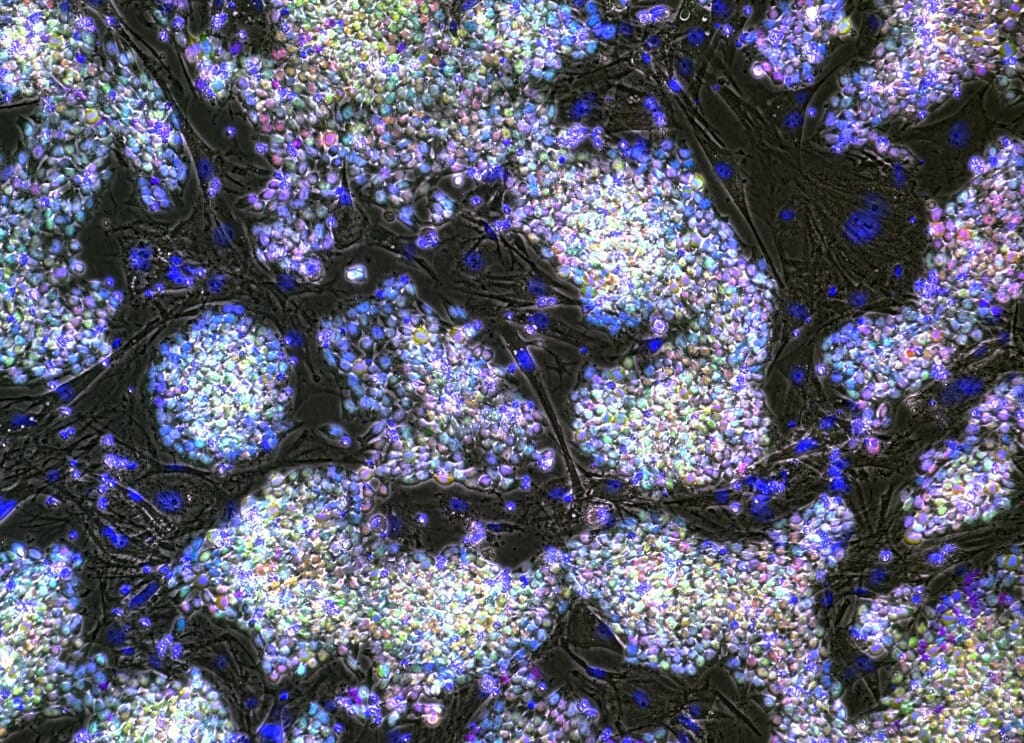
Stem cells derived from skin cells of a rhesus macaque monkey appear like multi-colored pearls atop spindly mouse cells that provide the stem cells with crucial support as they grow. These stem cells — which have the potential to become any type of cell in the body, but carry a genetic mutation linked to frontotemporal dementia —will be coaxed to form brain cells and used to study the development of the disease.
Julia Gambardella,
graduate student, Cellular & Molecular Pathology and Wisconsin National Primate Research Center;
John Maufort,
scientist, Morgridge Institute for Research and Wisconsin National Primate Research Center;
Marina Emborg,
professor, Medical Physics and Wisconsin National Primate Research Center
Digital-inverted microscope
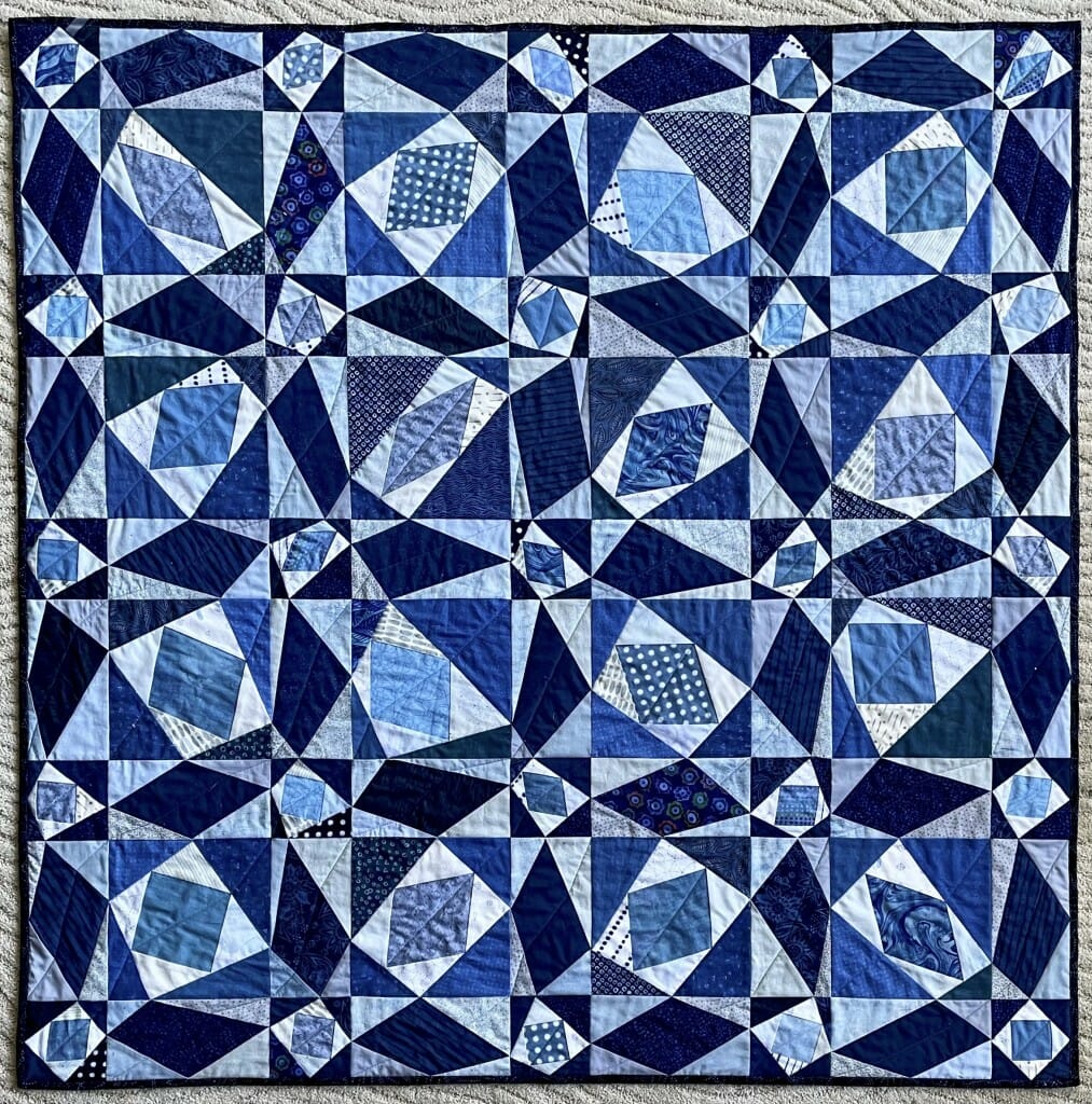
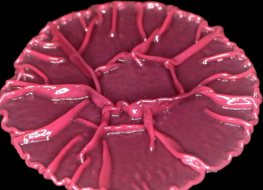
Follow this mathematical thread: The perfect regularity and symmetry of the traditional “Storm at Sea” patchwork has been disrupted in this quilt by shifting, in tandem, neighboring corners of the four-sided shapes in white and navy blue, placing the pairs of offset corners at a random position along the edge of the square or rectangle in which the quadrilaterals are inscribed. The 41 light blue quadrilaterals connect the midpoints of the edges of the white asymmetric quadrilaterals, and each is a visual illustration of Varignon’s Theorem, which proves that its shape must be a parallelogram.
Amy Wendt,
professor, Electrical and Computer Engineering
machine pieced and quilted cotton fabric
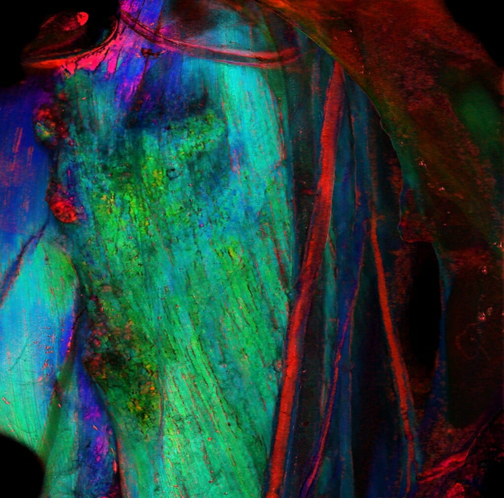
The strong, vertical line of the vagus nerve — appearing red with other nerves — carries motor and sensory information through the green muscle in a rat’s neck. Using a polarizing filter, researchers analyze the way peripheral nerves and their fatty layer of insulation reflect light. The technique may one day be used to help surgeons work around delicate nerves by distinguishing them from other tissue.
Rex Chin-Hao Chen,
graduate student, Biomedical Engineering;
Bruce Knudsen,
researcher, Biomedical Engineering;
Matthew LaLuzerne,
undergraduate student, Mechanical Engineering;
James Trevathan
scientist, Biomedical Engineering;
Kip Ludwig, professor,
Biomedical Engineering
Polarization Sensitive Optical Coherence Tomography
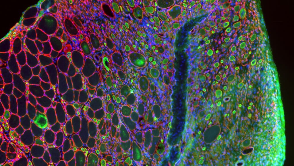
Tissue from a damaged muscle in the leg of a mouse includes healthy, uninjured cells appearing empty and outlined in red. Researchers studying the way aging and disease disrupt muscle growth and regeneration are interested in the difference between those healthy cells and the green-and-blue muscle stem cells, called satellite cells, which are missing a protein necessary for the healing process.
Jamie Hibbert,
postdoctoral fellow, Comparative Biosciences
Keyence microscope
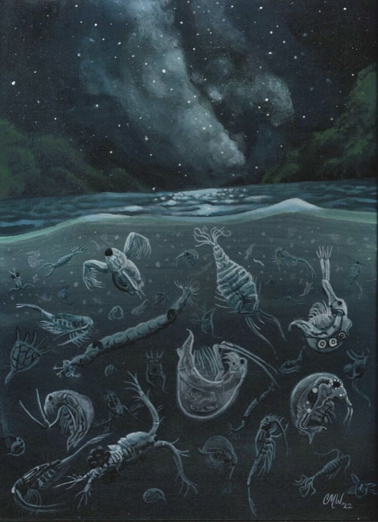
Some zooplankton — tiny swimming organisms that play an outsized role in the food webs of lakes — migrate daily up and down in the water column to find food and avoid predators. This painting, created by the artist during her residence at a UW–Madison limnological research station, depicts the migration of zooplankton species found in Escanaba Lake in Vilas County, Wisconsin.
Christina Weatherford,
science communications intern, Trout Lake Station
acrylic paint
This community of the bacteria Pseudomonas aeruginosa, a common cause of dangerous hospital-acquired infections, bands together into a biofilm to facilitate communication, stick to surfaces like medical devices and defend against antibiotics. Researchers study the P. aeruginosa genes relevant to biofilm formation in hopes of finding ways to disrupt them and kill the bacteria.
William Heelan,
graduate student, Pharmacy;
Ryan Ward,
graduate student, Genetics;
Amy Banta,
research scientist, Pharmacy;
Jason Peters
professor, Pharmacy
digital microscope
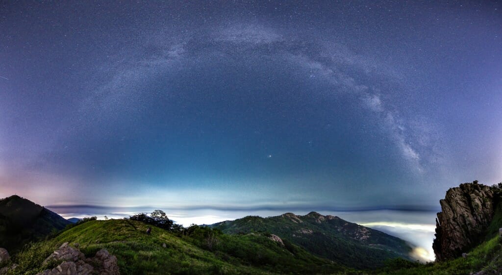
While it is difficult to see the stars of the Milky Way anywhere near the lights of a city, the galactic display in this assemblage of images taken above the Qingdao, China, area (population 7 million) is visible thanks to the blanket of fog dampening Qingdao’s light pollution.
Yingshun Sun,
graduate student, Atmospheric and Oceanic Sciences
digital camera
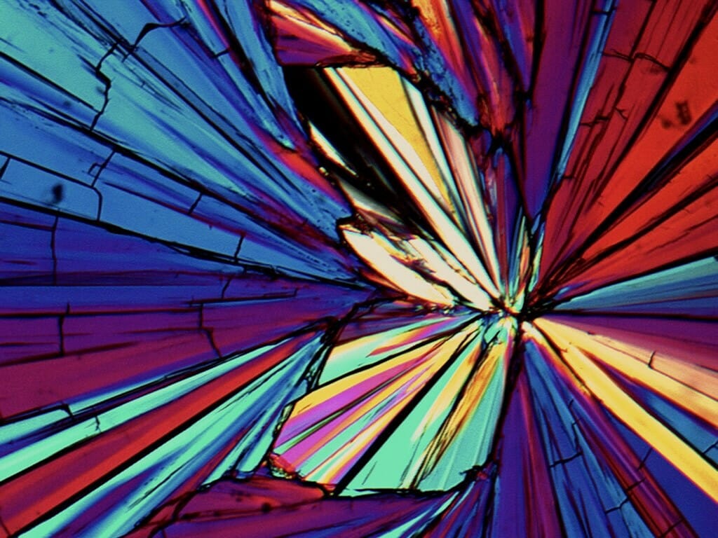
A polarizer makes arabitol shine as light passes through the sugar alcohol, illuminating its crystalline structure like a pane of stained glass or the sections of a butterfly wing. The crystal structures of drugs have profound influence on the way pharmaceuticals work in the body. Observing crystal formation in arabitol and similar substances helps researchers ensure safe and effective pharmaceuticals (and sharp LCD screens and perfectly tempered chocolate).
Amy Neusaenger,
graduate student, Pharmacy
polarized light microscope
Amy Neusaenger
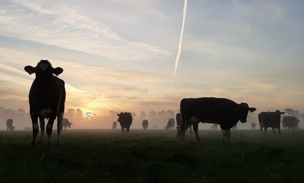
Dairy cows graze as the sun rises over a spring pasture in Ireland. Conor Holohan visited UW–Madison from University College Dublin to research ways to improve the nutrition and production of grass-fed cows on dairy farms in the U.S. Midwest.
Conor Holohan,
visiting researcher, Animal & Dairy Sciences
digital camera
Even from orbit 22,200 miles above Earth, the GOES-17 satellite captured the shockwave from the explosive eruption of the Hunga Tonga volcano as it passed through atmospheric water vapor over the Pacific Ocean on Jan. 15, 2022 — eventually circling the planet many times. The images were created from satellite data using software developed at UW–Madison in the 1970s and still in use worldwide.
Timothy J. Schmit, meteorologist, James P. Nelson III, data engineer, and Mathew M. Gunshor, researcher, all of the Cooperative Institute for Meteorological Satellite Studies
Digital rendering of satellite data
Continued from above gallery
The Cool Science Image Contest recognizes the technical and creative skills required to capture and create images, videos and other media that capably reveal something about science or nature while also leaving an impression with their beauty or ability to induce wonder. The contest is sponsored by Madison’s Promega Corp., with additional support from UW–Madison’s office of University Communications.
Winning entries are shared widely on UW–Madison websites, and all entries are showcased at campus science outreach events and in academic and lab facilities around campus throughout the year. See last year’s winners.
The contest judges were:
Steve Ackerman, professor of atmospheric and oceanic sciences and vice chancellor for research and graduate education
Kevin Eliceiri, director, Laboratory for Optical and Computational Instrumentation
Michael King, visual communications specialist, College of Agricultural and Life Sciences
Steve Paddock, former scientist, Molecular Biology
Kara Rogers, science writer and editor, Encyclopedia Britannica
Ahna Skop, professor of genetics
Kelly Tyrrell, director of media relations and strategic communications, University Communications
Craig Wild, videographer, University Communications
See more photo stories-
Interwoven
How Indigenous knowledge and science can work together to communicate about climate.
-
UW–Madison’s reach throughout Wisconsin adds up to $38.9 billion a year
The university supports more than 287,000 jobs and helps drive private-sector growth.
-
‘Being part of something bigger than you’ with Badger Volunteers
UW–Madison students head into the local community to find purpose and make a difference.
-
UW–Madison’s Tech Exploration Lab: Where the classroom meets the real world
The lab is built around a simple expectation: Students come to build, test and refine projects with real problems in mind.


