
Photo gallery The 2021 winners: Cool Science Image Contest
Ten images and two videos created by University of Wisconsin–Madison students, faculty and staff have been named winners of the 2021 Cool Science Image Contest.
A panel of nine experienced artists, scientists and science communicators judged the scientific content and aesthetic and creative qualities of scores of images and videos entered in the 11th annual competition. The winning entries showcase animals and plants, the invisibly small structures all around us, and stars and nebulae millions of millions of miles away.
An exhibit featuring the winners is open to the public at the McPherson Eye Research Institute’s Mandelbaum and Albert Family Vision Gallery on the ninth floor of the Wisconsin Institutes for Medical Research, 1111 Highland Ave., through December. A reception — open to the public — for the contest entrants will be held at the gallery on Oct. 7 from 4:30 to 6:30 p.m.
Winning submissions were created with point-and-shoot digital cameras, cutting-edge microscopes, and telescopes of both the backyard and mountaintop variety.
Because sometimes, there’s no substitute for the visual.
“An image often can convey meaning more effectively than words,” says Ahna Skop, a longtime contest judge, artist and UW–Madison professor of genetics and active ambassador for science. “We know from marketing and education research that adding a picture with words to a slide increases retention of knowledge by 65 percent. The visual communication of science is critical for the transference of knowledge broadly.”
Story continues after gallery
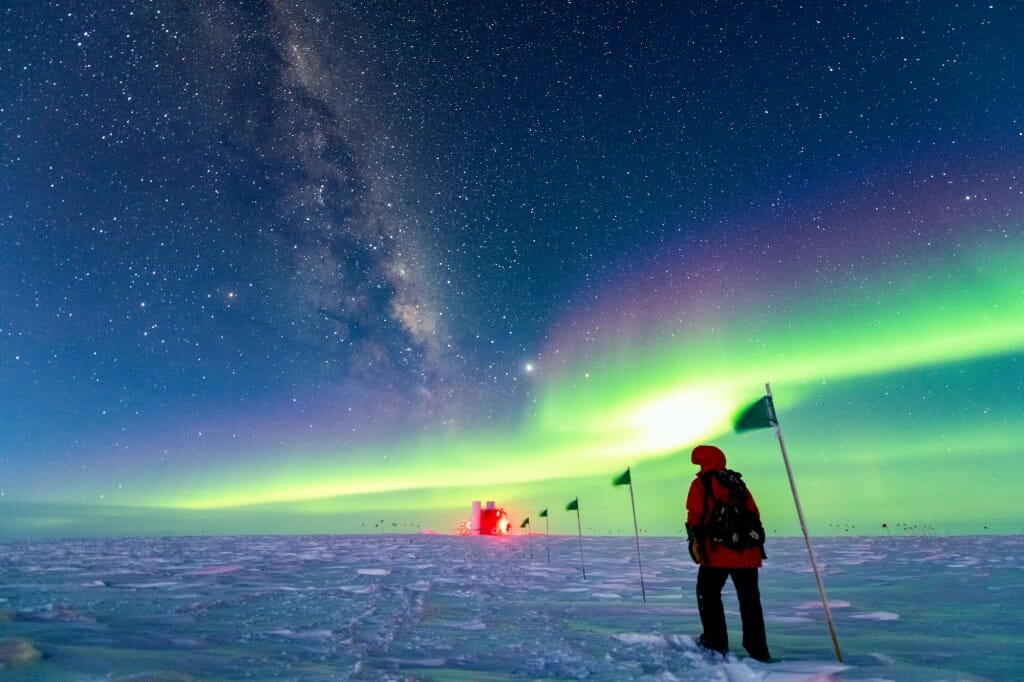
A “winterover” — one of the two staff members who stay through the minus-100-degree Fahrenheit nights of Antarctica’s coldest months — hikes underneath the stars and aurora to the South Pole home of IceCube, a UW–Madison-led neutrino telescope, in the 2021 Cool Science Image Contest winning photo. Yuya Makino
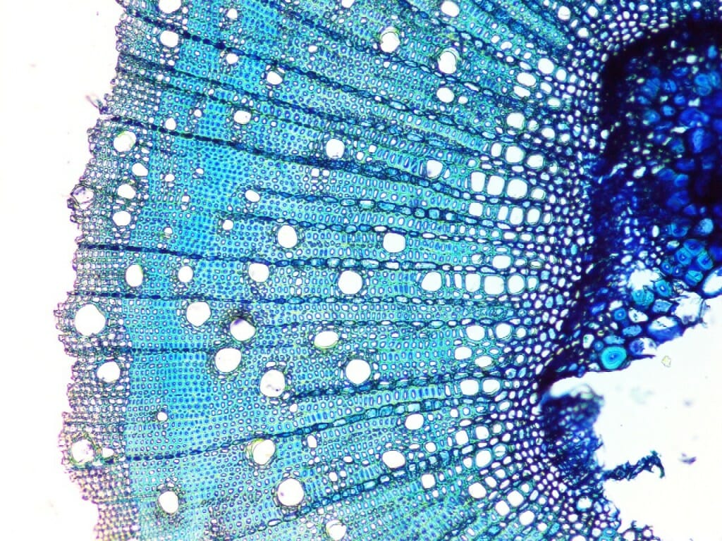
The large holes in this cross-section of a stalk of desert stringybark, Eucalyptus arenacea, are conduits through the plant tissue that help researchers quantify the way the plant — native to dry parts of Australia — adapts to a new, wetter environment.
Kennah Konrad,
undergraduate student, Botany;
Duncan Smith,
assistant scientist, Botany
compound microscope
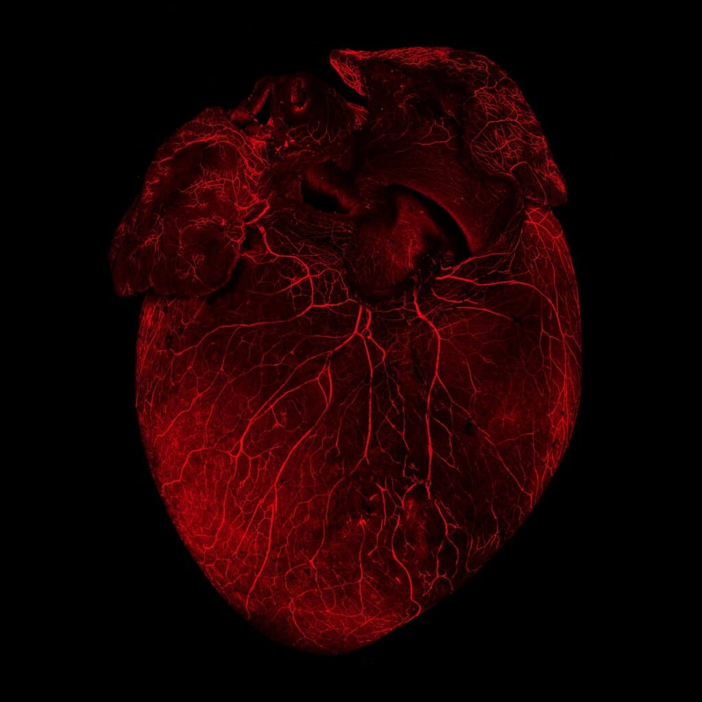
Fluorescent antibodies highlight the extensive nervous system of a mouse heart. By creating maps of cardiac nerves with unprecedented accuracy, researchers can explore how those nerves influence heart function.
Rebecca Salamon,
graduate student, Cell and Regenerative Biology
confocal microscope
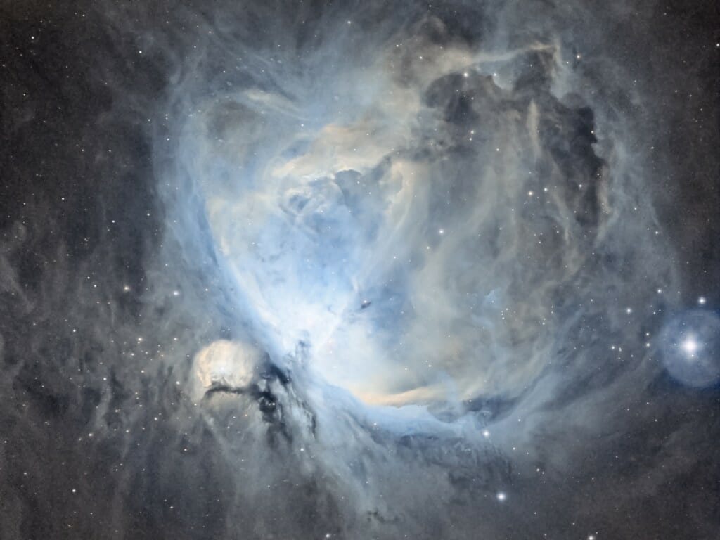
Messier 42, known as the Orion Nebula, is in the sword of the constellation Orion and is one of the brightest nebulae in the sky. At just 1,400 light-years away and 24 light-years across, it is one of the closest and largest regions of dense gas and dust in which stars are formed.
Jeffrey E. Shokler,
associate director, Office of Undergraduate Advising
refractor telescope and CCD camera
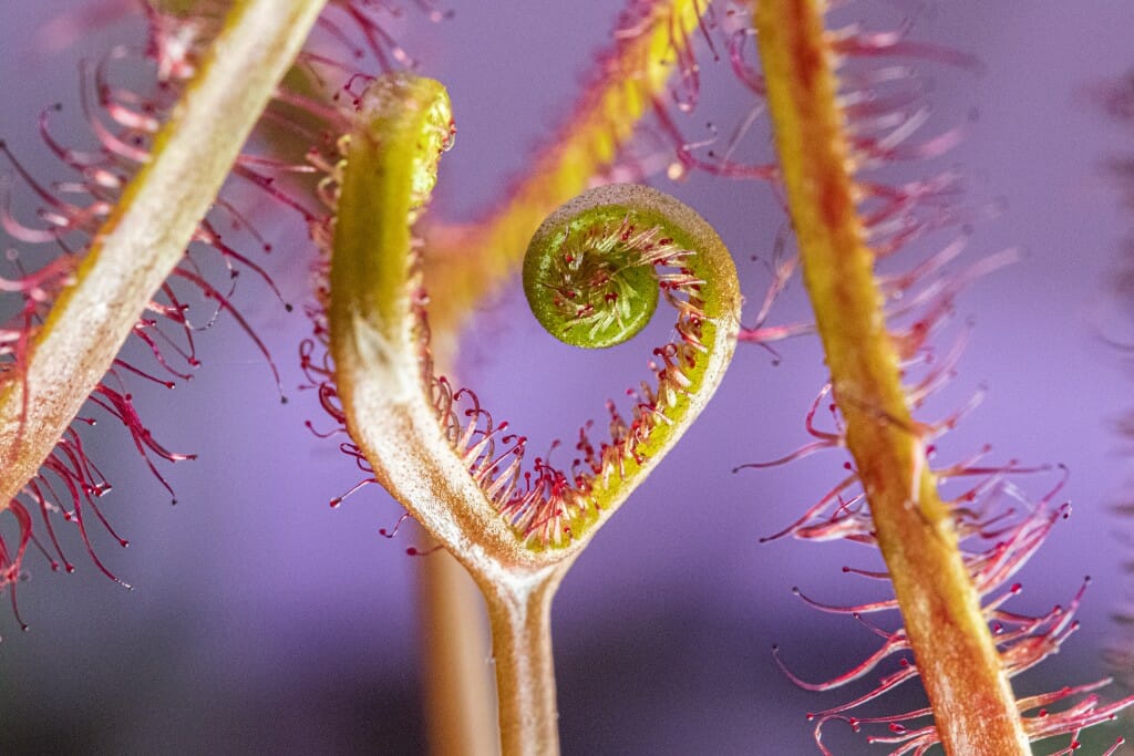
The carnivorous sundew plant snags insect meals with armloads of tentacles that it can move to tighten its grip and bog down prey in sticky secretions. The leaves roll up around a meal to facilitate digestion by enzymes and absorption of the nutrients.
Nisha Iyer,
postdoctoral fellow, Wisconsin Institute for Discovery
digital camera
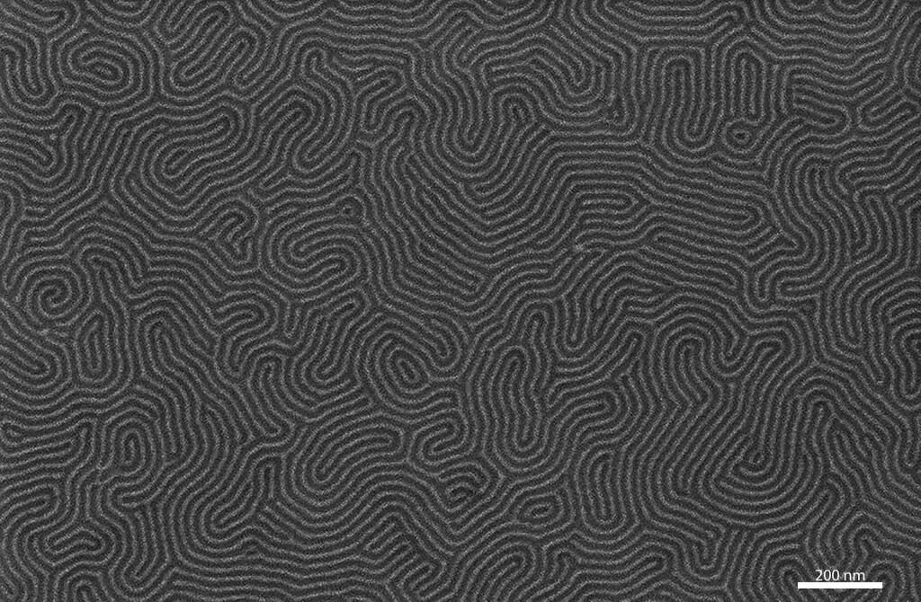
Mazes of tiny structures less than 15 billionths of a meter across and made of some of the smallest ribbons of graphene — layers of carbon just a single atom thick — ever fabricated represent an important step toward graphene-based telecommunications devices.
Joel Siegel and Margaret Fortman,
graduate students, Physics;
Jian Sun,
graduate student, Materials Science;
Jonathan Dwyer,
PhD alumnus, Chemical Engineering
scanning electron microscope
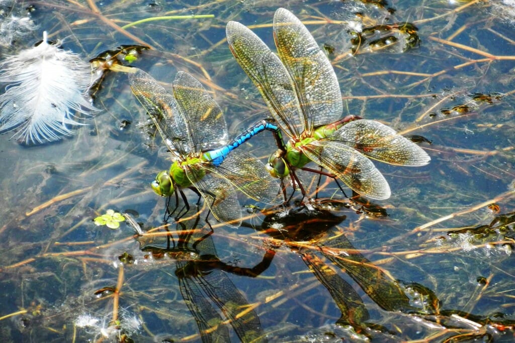
A pair of mating dragonflies pause on the surface of a Minnesota pond. Dragonfly coupling begins with the male (with blue markings) gripping the female with claspers at the very end of his abdomen. To complete the act, the female will bend her abdomen underneath her body to meet the male’s abdomen and create a characteristic heart shape.
Shin-Tsz (Lucy) Kuo
undergraduate student, Computer Science and Economics
digital camera
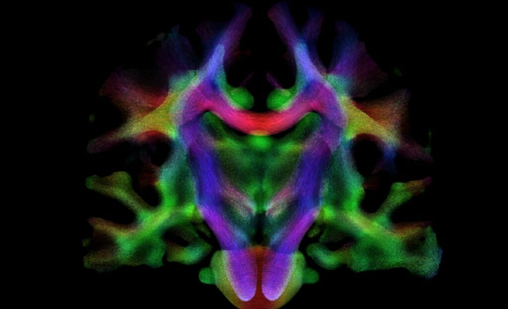
White matter, the connective nerve tissue of the brain, has been colored according to the predominant orientation of fibers — red, right-left; green, front-back; blue, up-down — in different regions of the human brain to reveal pathways traversing the regions. Understanding white matter organization may offer insights into normal brain development as well as into the study of neurological disorders.
Jose Guerrero,
postdoctoral fellow, Medical Physics;
Andrew Alexander,
professor, Medical Physics;
Peter Ferrazzano
professor, Pediatrics
magnetic resonance imaging scanner
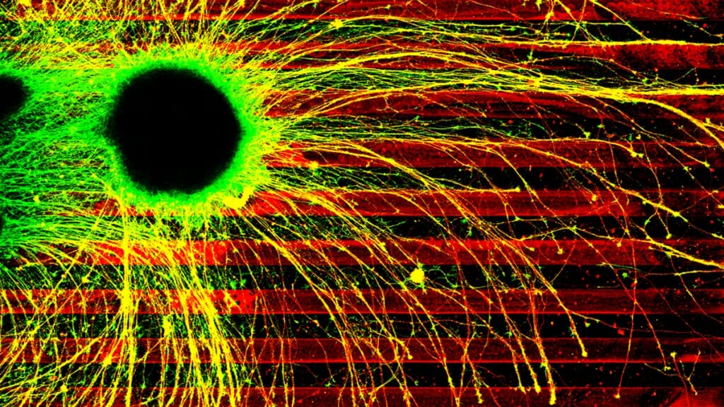
The yellow connecting arms, called axons, of diseased human brain cells grow willy-nilly across boundaries of inhibitory chemicals (the red stripes). Healthy axons would precisely follow the dark lanes, giving researchers the opportunity to test the effects of disease-causing mutations on axon growth.
Timothy Catlett
graduate student, Cell and Molecular Biology;
Timothy Gomez,
professor, Neuroscience
confocal microscope
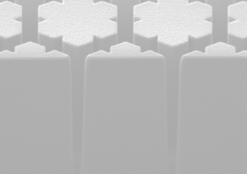
By varying the exact size and shape of these micrometer-wide, star-shaped pillars etched into a silicon wafer, researchers can carefully manipulate light passing through a lens to correct for aberrations that would otherwise focus different wavelengths of light on different points in space.
Gregory Holdman,
graduate student, Physics
focused ion beam and scanning electron microscope
Recurrent neural networks are the computing engines behind state-of-the-art applications from self-driving cars to speech recognition like Amazon’s Alexa. The behavior of these networks is challenging to characterize, but it can be visualized for small networks. This video displays the behavior of a network with just three neurons, showing the way their output evolves by mapping their values in blue. The result, a fractal structure called a “strange attractor,” could help researchers better understand the behavior and characteristics of these kinds of networks.
David J. Nowak, alumnus and auditing student; Robert D. Nowak, professor, Electrical and Computer Engineering
Captured at 20,000 frames per second, this video shows the shock-wave-induced mixture of two gasses — raw imagery on the left; adjusted to better reflect concentration of the lighter gas on the right. Experiments like this are run in the 9-meter-tall Wisconsin Shock Tube, depicted at left, to simulate and explore mixing at the interface of materials in extreme conditions like nuclear fusion, supernovae and hypersonic propulsion.
Josh Herzog, postdoctoral fellow, and Professor David Rothamer, both of Mechanical Engineering; Riccardo Bonazza, professsor, Engineering Physics
Continued from above gallery
There can be an ineffable sort of something that makes a particularly effective science image — it’s the “Cool” in Cool Science Image Contest — but the good ones have much in common.
“You’ll know it when you see it. It’s like seeing “Starry Night” or the “Mona Lisa” for the first time, in person. They hit you deep and quickly,” Skop says. “They are beautiful to the eye, simple, and convey meaning. Some images just take your breath away. Looking deeper they exquisitely communicate the secrets of science beautifully.”
The Cool Science Image Contest recognizes the technical and creative skills required to capture images or videos that capably reveal something about science or nature while also leaving an impression with their beauty or ability to induce wonder. The contest is sponsored by Madison’s Promega Corp., with additional support from the UW–Madison Division of the Arts.
Winning entries are shared widely on UW–Madison websites, and all entries are showcased at campus science outreach events and in academic and lab facilities around campus throughout the year. Because there was no opportunity to show off the 2020 contest winners in-person, this year’s exhibit is a double-feature for both the 2020 and 2021 contests. See last year’s winners.
The contest judges were:
Steve Ackerman, professor of atmospheric and oceanic sciences and vice chancellor for research and graduate education
Terry Devitt, emeritus director of research communications, University Communications
Kevin Eliceiri, director, Laboratory for Optical and Computational Instrumentation
Michael King, visual communications specialist, College of Agricultural and Life Sciences
Steve Paddock, former scientist, Molecular Biology
Kara Rogers, science writer and editor, Encyclopedia Britannica
Ahna Skop, professor of genetics
Kelly Tyrrell, director of research communications, University Communications
Craig Wild, videographer, University Communications
See more photo storiesTags: recent sightings
-
Interwoven
How Indigenous knowledge and science can work together to communicate about climate.
-
UW–Madison’s reach throughout Wisconsin adds up to $38.9 billion a year
The university supports more than 287,000 jobs and helps drive private-sector growth.
-
‘Being part of something bigger than you’ with Badger Volunteers
UW–Madison students head into the local community to find purpose and make a difference.
-
UW–Madison’s Tech Exploration Lab: Where the classroom meets the real world
The lab is built around a simple expectation: Students come to build, test and refine projects with real problems in mind.


