
Photo gallery The winners: Cool Science Images 2018
Ten images and two videos by University of Wisconsin–Madison students, faculty and staff have been named winners of the 2018 Cool Science Image Contest.
A panel of eight experienced artists and scientists judged the scientific content and aesthetic and creative qualities of 171 images and videos, a record number of entries for the eighth annual competition. [story continues after gallery]
2018 Winning Cool Science Images
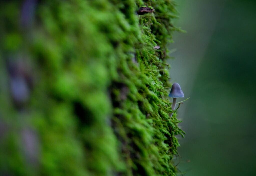
A solitary Mycena mushroom grows among moss on the bark of an oak tree in Wisconsin’s Yellowstone State Park.
Francisco Barros, graduate student, cell and molecular biology, Huttenlocher Lab, Department of Pediatrics; digital camera
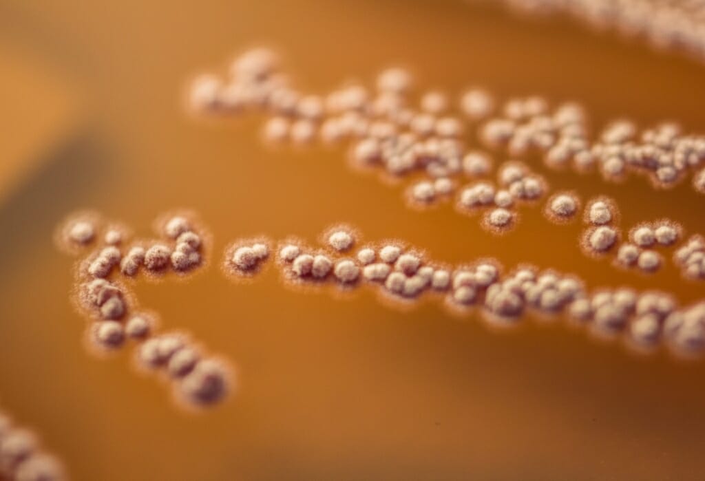
Bacteria from a Costa Rican parasitoid wasp grow on a culture plate. The bacteria were prospects in UW–Madison researchers’ search for new antimicrobial compounds.
Caitlin Carlson, research specialist, Currie Lab, Department of Bacteriology; digital camera
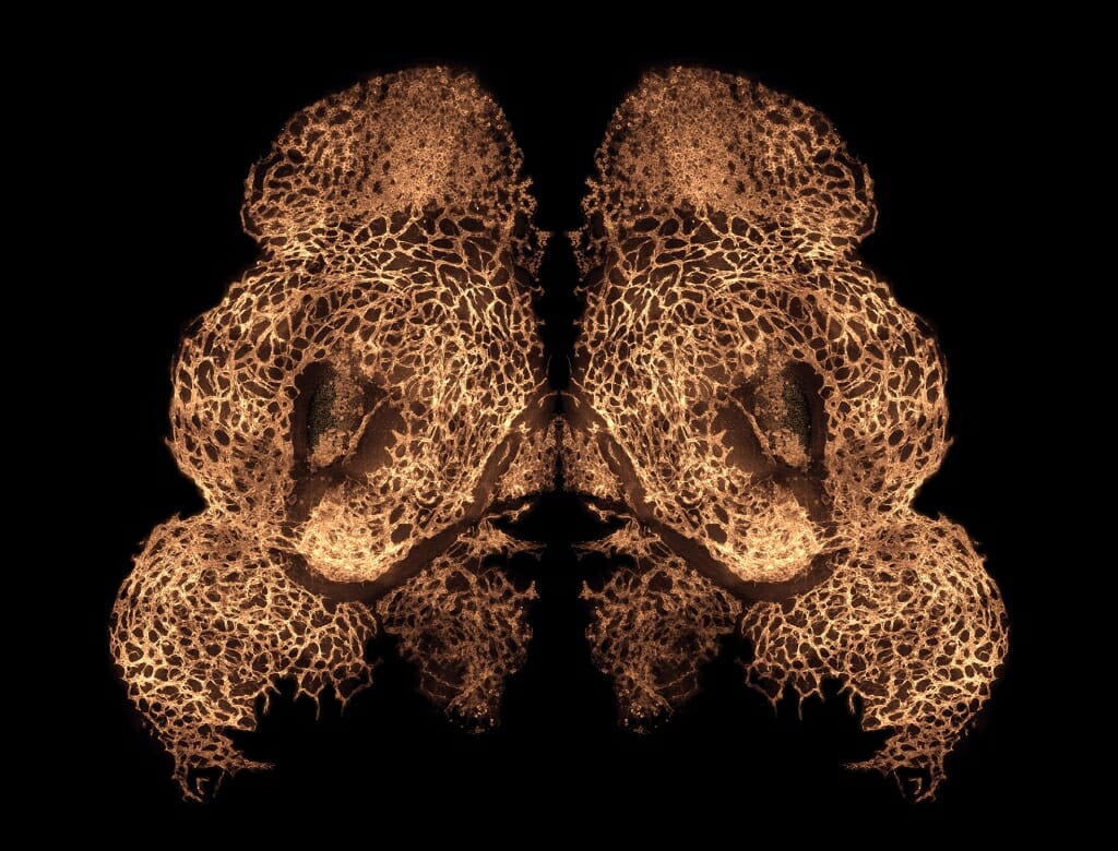
The blood vessels developing in the face of an 11-day-old mouse embryo are most concentrated in areas that will become the upper lip, while the two empty spots will become nostrils. Facial development in mice and people is so similar, this is much what a human embryo’s facial blood vessels look like at 34 days.
Hannah Chung, graduate student, Department of Comparative Biosciences; confocal microscope
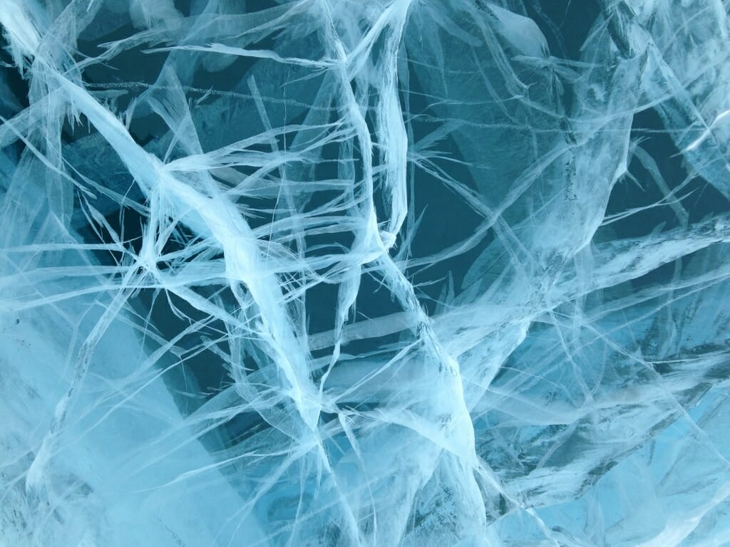
The ice on Antarctica’s Lake Vanda is 10 feet thick and particularly clear. Lake Vanda’s liquid water is also salty and particularly warm — about 70 degrees in the 70-meter depths — suggesting salty groundwater is seeping in. Researchers believe studying Lake Vanda may help them in a search for subsurface water on Mars.
Hilary Dugan, integrative biology professor, Center for Limnology; smartphone camera
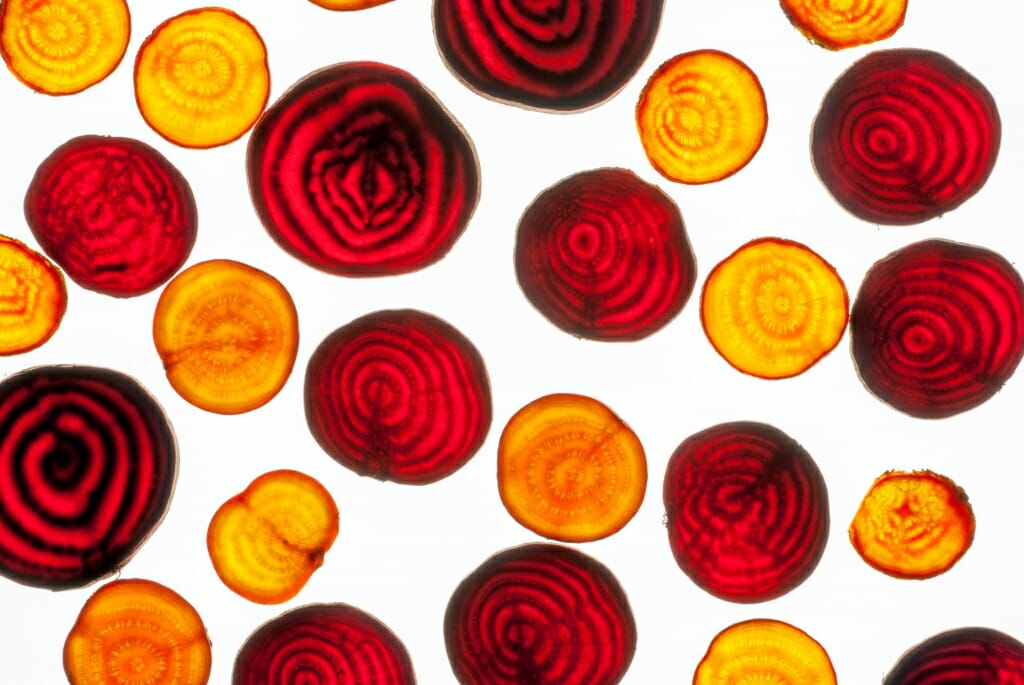
Thin slices of beets on a light table show off bands of red and yellow. UW–Madison botanists are studying how the pigments that create the vivid colors evolved in a very small group of plants.
Sarah Friedrich, media specialist, Department of Botany; digital camera
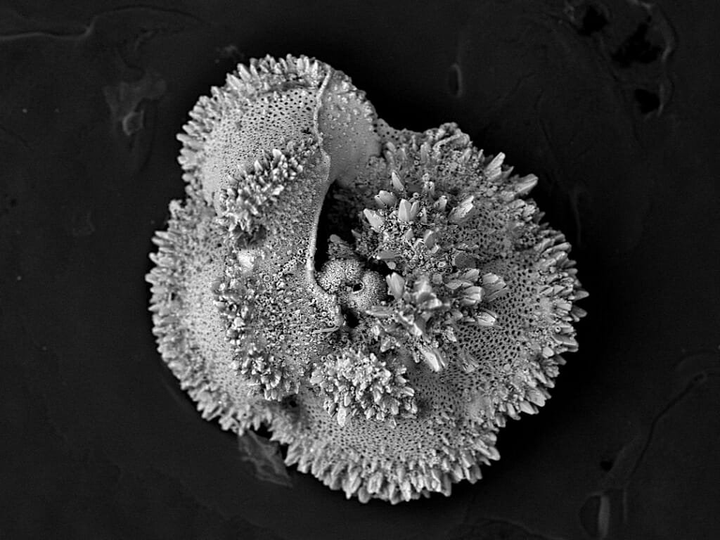
This fossil marine zooplankton, of the 55-million-year-old foraminifer species Morozovella velascoensis, is just 400 microns across — about one-third the width of a grain of sand. But chemical analysis of the tiny fossils can help researchers understand Earth’s climate during a warming period similar to today.
Brittany Hupp, graduate student, Kelly Lab, Department of Geosciences; scanning electron microscope
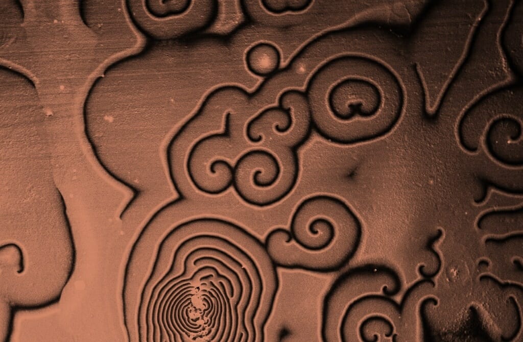
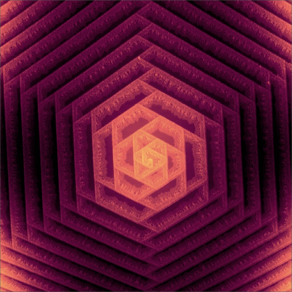
These spiral patterns formed spontaneously when indium and cobalt were mixed and deposited as an electrode for use in biomass conversion reactions. Metal alloys can have qualities that make them better catalysts than their individual components alone.
Stephen Kubota, graduate student, Department of Chemistry;
scanning electron microscope
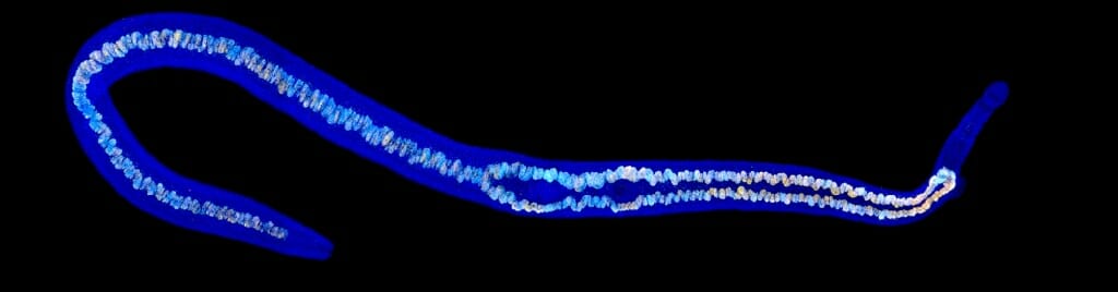
The mixed colors running the length of this otherwise blue-stained parasitic flatworm Schistosoma mansoni allow researchers to visualize the expression of various genes in its gut. The parasite causes the disease schistosomiasis in hundreds of millions of people each year, and understanding the development of their guts might provide a target for treatment.
Jayhun Lee, postdoctoral fellow, Newmark Lab, Morgridge Institute for Research;
laser scanning confocal microscope
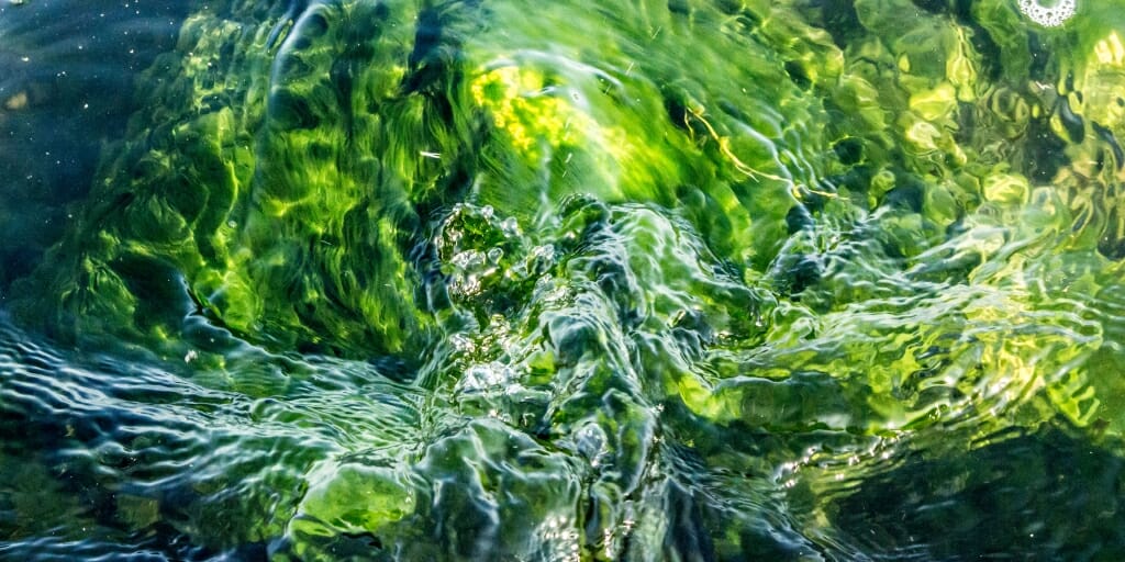
Algae and water in Lake Monona ripple in sunlight. UW–Madison researchers study the way runoff from farm fields and invasive species contribute to conditions ideal for algae blooms.
Rhianna Miles, digital media specialist, Center for Sustainability and the Global Environment; digital camera
The complex spiral patterns in the nanostructure of tungsten diselenide look and work as a fingerprint, identifying the arrangement of atoms within the material. Understanding the arrangements and how they form could broaden the material’s use in applications from solar energy to computer memory.
Melinda Shearer, graduate student, Hamers and Jin labs, Department of Chemistry; atomic force microscope
Video Winners
While tracking lions to observe hunting behavior in Rwanda’s Akagera National Park, University of Wisconsin–Madison researchers came across an impala carcass. They set up a hidden camera to see if the male lions were willing to scavenge food. They got their answer.
Drew Bantlin, graduate student, Nelson Institute for Environmental Studies
Early in the development of a zebrafish, sensory neurons extend from the spinal cord and across both sides of the embryo to allow it to sense touch. In this video clip, representing 24 hours of growth captured by light sheet microscopy, sensory neurons along both sides of the spinal cord show up as two bright lines. Branching threads of nerve cells, called axons, grow out from the cords.
Jiaye “Henry” He, of the Morgridge Institute for Research, and Liz M. Haynes, postdoctoral fellow in Integrative Biology
The number and quality of the submissions are evidence that researchers understand pictures are a great way for their work to reach a wide audience, according to the contest judges.
“Beauty drives curiosity for me, as a scientist. It’s the same for people who are curious, but not scientists,” says Ahna Skop, a contest judge and UW–Madison professor of genetics. “When people see a good image you’ve made, they say, ‘That looks amazing. Tell me what it is.’”
The 2018 winners were made with intricate, high-tech tools over the course of days of capturing and processing, with smartphone cameras pulled from pockets to catch a moment of serendipity, and with all manner of methods in-between.
The subjects are plant, animal and mineral, pictured from a distance — a safe distance, in the case of one carnivorous king of the jungle — down to the graceful nanoscale arrangement of metallic molecules.
“It’s all data, on some level, but it’s often beautiful,” says Skop, who grew up in a house full of artists, and creates her own art in and out of the lab. “As a microscopist, we work to visualize things you can’t see with the human eye. And for many of us, our technical skill and how we see this mysterious and beautiful world influences how we frame and capture the image. And more often than not we discover works of art.”
The Cool Science Image Contest reflects those discoveries and recognizes the technical and creative skills required to capture images or video that document science or nature. The contest is sponsored by Madison’s Promega Corp., with additional support from DoIT Digital Publishing and Printing Services and the UW–Madison Arts Institute.
Winning entries are shared widely on various UW–Madison websites, and all entries are showcased in a slide show at the Wisconsin Science Festival and in concert with a fall exhibit of winners at the McPherson Eye Research Institute’s Mandelbaum and Albert Family Vision Gallery.
UW–Madison 2018 Cool Science Image Contest winners are:
Drew Bantlin, graduate student in the Nelson Institute for Environmental Studies, for a trail camera video of an African lion sneaking up to and absconding with the carcass of an impala.
Francisco Barros, graduate student in cell and molecular biology, for his photo of a solitary Mycena mushroom growing among moss on the bark of an oak tree in Wisconsin’s Yellowstone State Park.
Caitlin Carlson, research specialist in Bacteriology, for her photo of bacteria colonies cultured from a Costa Rican parasitoid wasp that may be prospects for new antimicrobial compounds.
Hannah Chung, graduate student in Comparative Biosciences in the School of Veterinary Medicine, for a micrograph of blood vessels developing in the face of an 11-day-old mouse embryo.
Hilary Dugan, integrative biology professor in the Center for Limnology, for a photo of ice on Antarctica’s Lake Vanda, a lake with a structure that may be similar to underground water deposits on Mars.
Sarah Friedrich, media specialist in Botany, for a photo of thin slices of red and yellow beets studied by geneticists tracking the evolution of their vivid pigments.
Jiaye “Henry” He, graduate student at the Morgridge Institute for Research, and Liz M. Haynes, postdoctoral fellow in Integrative Biology, for their video of the development of sensory neurons extending from the spinal cord of an embryonic zebrafish.
Brittany Hupp, graduate student in Geosciences, for a scanning electron micrograph of 55-million-year-old fossilized marine zooplankton used to study climate change.
Stephen Kubota, graduate student in Chemistry, for a scanning electron micrograph of spiral patterns formed when indium and cobalt were mixed to make an electrode for catalyzing biomass conversion reactions.
Jayhun Lee, postdoctoral fellow at the Morgridge Institute for Research, for an image from a laser scanning confocal microscope of gene expression in the gut of a disease-causing parasitic flatworm.
Rhianna Miles, digital media specialist in the Center for Sustainability and the Global Environment, for a digital camera image capturing the sunlit rippling of water and algae in Lake Monona.
Melinda Shearer, graduate student in Chemistry, for an image taken with an atomic force microscope of fingerprint patterns that reveal the arrangement of atoms in the nanostructure of tungsten diselenide.
The judges were:
Steve Ackerman, UW–Madison professor of atmospheric and oceanic sciences
John Baldacchino, director, UW–Madison Arts Institute
Kevin Eliceiri, director, UW–Madison Laboratory for Optical and Computational Instrumentation
Hyunsoo Léo Kim, photographer, University Communications
Steve Paddock, associate scientist, UW–Madison Department of Molecular Biology
Paula Panczenko, director, Tandem Press
Kara Rogers, science writer and editor, Encyclopedia Britannica
Ahna Skop, UW–Madison professor of genetics
Tags: arts, awards, faculty and staff, science, students
-
Interwoven
How Indigenous knowledge and science can work together to communicate about climate.
-
UW–Madison’s reach throughout Wisconsin adds up to $38.9 billion a year
The university supports more than 287,000 jobs and helps drive private-sector growth.
-
‘Being part of something bigger than you’ with Badger Volunteers
UW–Madison students head into the local community to find purpose and make a difference.
-
UW–Madison’s Tech Exploration Lab: Where the classroom meets the real world
The lab is built around a simple expectation: Students come to build, test and refine projects with real problems in mind.


