Science + art exhibit focuses on the beauty of a cure
An unusual exhibit focusing on cancer recovery through the lens of art and science has opened in the Biochemistry Department.
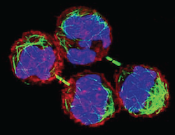
Cells are shown just after completing division in one of the works in the exhibit “Bioimaging: Domains and Dimensions,” opening Feb. 22 at the Biochemistry Addition. The green bundle between the pairs of daughter cells helps them separate. Image by Ahna Skop.
One goal of “Bioimaging: Domains and Dimensions: Healing through Art and Science: Cancer” is “to do a better job of showing the public what kind of science has gone on, and is going on, at Wisconsin,” says co-organizer Dave Nelson, professor emeritus of biochemistry.
An artwork by Madison artist Carol Bjerke that stretches 110 feet across one wall is her effort to make sense and meaning of her experience battling and surviving colon cancer. “Every three days while changing my ostomy appliance, I drew the circle of sealant and then photographed the prepared piece before placing it over my stoma” says Bjerke. “At the end of the year l had one hundred and twenty-two images to mark the rhythm of my existence.”
Images on the opposite wall highlight groundbreaking cancer research at UW–Madison and local biomedical companies. At McArdle Laboratory in the late 1940s, for example, Charles Heidelberger developed a method to label carcinogenic compounds with radioactive isotopes made available during the Manhattan Project that developed the atomic bomb.
“Heidelberger followed the compounds through healthy cells and discovered that the carcinogens bound to specific proteins; this was one of the first clues to how these agents work,” says Nelson. In the 1950s, Heidelberger identified a compound called 5FU that is still used to treat colon cancer.
Another set of images concentrates on the inherent beauty of scientific images. “We are including images generated in labs showing cancer cells, made with bright fluorescent probes,” says exhibit coordinator Olga Trubetskoy. “You can view them as pieces of art, but each is accompanied by a plain-language description of the scientific significance.”
The modern images also include a 3-D model of a protein that is the main target of 5FU. “What we hope people will see is that there is actually an artistic element to science, and that they have a lot to be proud of in their university,” says Nelson.
Future exhibits in “Bioimaging: Domains and Dimensions” will focus on model organisms like the fruit fly that are instrumental to biology and medicine. This fall, Nelson and Trubetskoy hope to bring “Beyond X-Rays,” a large exhibit from the Boston Museum of Science focused on technology such as MRI and CT scanning, another field of intense activity by UW–Madison researchers.
Mixing art, science and disease is not quite as strange as it sounds, says Nelson. “This exhibit is not just about cancer, it’s about recovery and survival; it’s about showing that the research done and exhibited here has contributed mightily to that survival.”
The exhibit will be open from 7:30 a.m. to 6 p.m. until March 31.
Tags: arts, biosciences, cancer