Astronomy technique could help assess deadly melanomas
As a young graduate student with a passion for surfing, Andy Sheinis soaked up a lot of California sun.

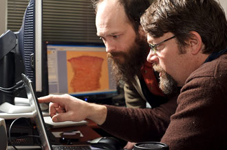
Andrew Sheinis (right), professor of astronomy, and Trevor Irwin (left), honorary associate / fellow in molecular biology, take readings from a microscope in the dermatology department. Sheinis, who is at high risk for skin cancer, is applying his knowledge of astrophysics and instrument-making ability to devising a new method for assessing skin blots that could become cancerous.
Photo: Bryce Richter
But because of his youthful pursuit, Sheinis, now a UW–Madison professor of astronomy, is at high risk for melanoma, skin cancer that can turn deadly if not detected and treated in its earliest stages.
“I’m one of those people who has to be checked every six months,” says Sheinis, who, in addition to his research on the hearts of distant galaxies, is also an expert instrument maker for ginormous telescopes such as those at Hawaii’s W.M. Keck Observatory and the South African Large Telescope (SALT). “About half the time I go in they have to cut something off.”
About 10 years ago, during a visit to the dermatologist, it struck Sheinis that the same techniques astronomers use to parse starlight could be used to assess the skin blots that may become cancerous. The system now used most commonly to evaluate worrisome moles is the human eye and the brain, explains Sheinis. Clinicians use the “ABCDE rule” — asymmetry, border irregularity, color, diameter and evolving — to assess the cancerous potential of moles.
“They don’t take pictures of them most of the time, which I found kind of surprising,” notes Sheinis. “The success of this technique depends strongly on the training and experience of the dermatologist.”
At about the time Sheinis began regular screening for melanoma, he was building a hyperspectral imager for the giant Keck Telescope. The technology, originally invented for remote-sensing applications by the Department of Defense, is a low-cost and speedy way to sample the spectrum of light in every pixel of an image and build a three-dimensional “data cube.” It is used in astronomy to tease information about the size and composition of celestial objects millions or billions of light years from Earth.
The technology, Sheinis notes, can be compressed into a device the size of a camera and with the help of his industrial partner, Bodkin Design and Engineering in Newton Mass, is now being integrated into a microscope at UW–Madison’s Laboratory for Optical and Computational Instrumentation (LOCI).
Sheinis, along with co-principal investigators Kevin Eliceiri, of molecular biology and biomedical engineering and director of LOCI and Sharon Weber, professor of surgery in the UW–Madison School of Medicine and Public Health, are conducting the study which is supported by a grant from the Wallace H. Coulter Translational Research Partnership program. The Coulter program supports translational research in biomedical engineering, in the UW–Madison Department of Biomedical Engineering.
In the context of assessing moles for their potential to become cancerous, the method, in a single snapshot, captures a three dimensional image in anywhere from 90 to several hundred wavelengths of light. It is quick, noninvasive, inexpensive and holds clinical promise not only for identifying worrisome moles but also for accurately mapping their extent, which is critical information when it comes to surgically excising them.
“A lot of skin cancer has ill-defined margins,” says Yaohui Gloria Xu, an assistant professor of dermatology in the UW–Madison School of Medicine and Public Health who is helping to test the technology’s clinical potential. “Sometimes you don’t really know where to cut.”
She explains that surgeons, while seeking to minimize the amount of tissue they remove, need to be sure they are getting all of the cancerous cells so that a melanoma does not return. “This could help us cut more accurately,” she says. “We are always chasing cancers.”
But the skin cancer assessment potential of the technology is just now being assessed using cancerous tissue, mounted on microscope slides, from about 50 patients as a first critical test being conducted in the dermatology lab of B. Jack Longley, professor of dermatology in the School of Medicine and Public Health. By comparing the data from the hyperspectral imager to the histology or microscopic anatomy of the mole, the gold standard for diagnosing melanoma, Sheinis, Xu and their colleagues will be able to test the accuracy of the method before trying the technology in a clinical setting.
“The idea is to take this device from astronomy and apply it to melanoma,” says Eliceiri. “This is very new stuff. We don’t know if it’s going to work, but that’s why we want to try it.”
Eliceiri explains that using hyperspectral imaging to gauge cancerous tissue isn’t a new idea, but that the technology Sheinis brings to the table captures more data by sampling a broader swath of the electromagnetic spectrum: “Andy’s device has more colors available. With those additional channels, we may get a better readout of the cancer.”
Elirceiri also points out that astronomers have decades of experience processing the huge amounts of data gathered by their spectral sampling of stars and galaxies, experience that may lend itself to biomedical applications of spectral imaging.
Xu, while optimistic about the technology, expresses the cautious perspective of the clinician: “The technology is in its infancy and the device isn’t going to persuade us right away, but it deserves a chance. It could sharpen our clinical judgment.”
