Photo gallery 2024 winners: Cool Science Image Contest
Winners in the University of Wisconsin–Madison’s 14th annual Cool Science Image Contest span a wide range of perception — from molecules interacting in the membranes of individual cells to the stretchy insides of a delicious cheese puff to a sky full of colors made by the strongest solar storm in decades.
A panel of experienced artists, scientists and science communicators chose 10 winning still images and two videos based on the aesthetic, creative and scientific qualities that distinguished them from this year’s field of submissions.
The winning entries showcase the research, innovation, scholarship and curiosity of the UW–Madison community through traditional fine art techniques used to study the physics of interacting liquids, the surprising and beautiful results of chemical and geological processes, and new ways to manipulate and reveal biological processes.
The winning images go on display next week in an exhibit at the McPherson Eye Research Institute’s Mandelbaum and Albert Family Vision Gallery on the ninth floor of the Wisconsin Institutes for Medical Research, 1111 Highland Ave. The exhibit, which runs through the end of 2024, opens with a reception — open to the public — at the gallery on Thursday, Oct. 3, from 4:30 to 6:30 p.m.
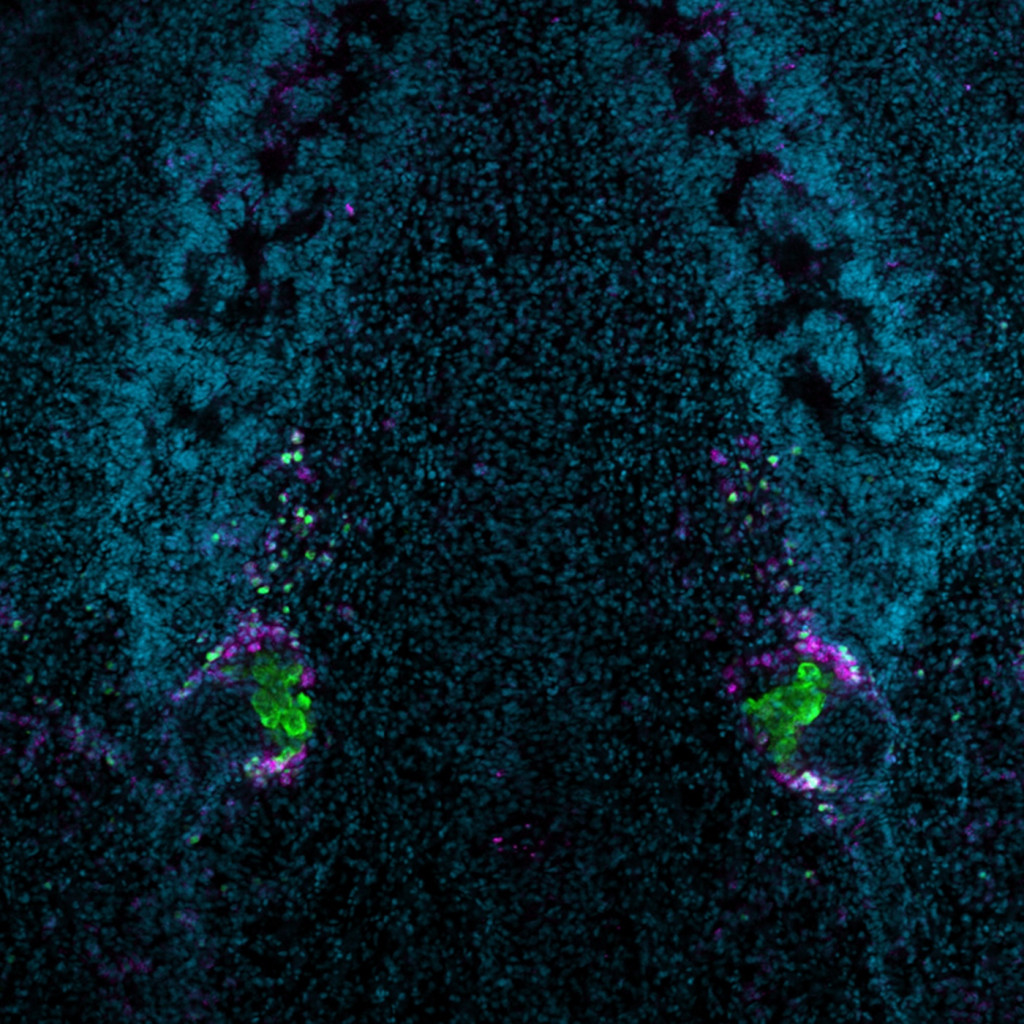
A planarian flatworm, Schmidtea mediterranea, can regrow a brain (the horseshoe shape of blue cell nuclei) and ovaries (purple and green cells) from a tail fragment even after losing its head! The Newmark/Issigonis Lab studies the biochemical signals key to the development of “germline” cells — the ones that pass on genetic information to offspring. Katherine Browder, graduate student, Integrative Biology Melanie Issigonis, research investigator, Morgridge Institute for Research Phil Newmark, investigator, Morgridge Institute for Research Laser scanning confocal microscope
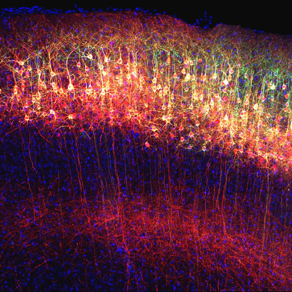
Neurons in a mouse can migrate among the outer layers of the brain, called the cortex. Researchers in Erik Dent’s lab label parts of the cells — greenish cell bodies and red axons (the branches the cells use to reach out to their neighbors) — to track the way shifting protein levels affect migration. Erik Dent, professor, neuroscience Kendra Taylor, graduate student, Neuroscience Scanning confocal microscope
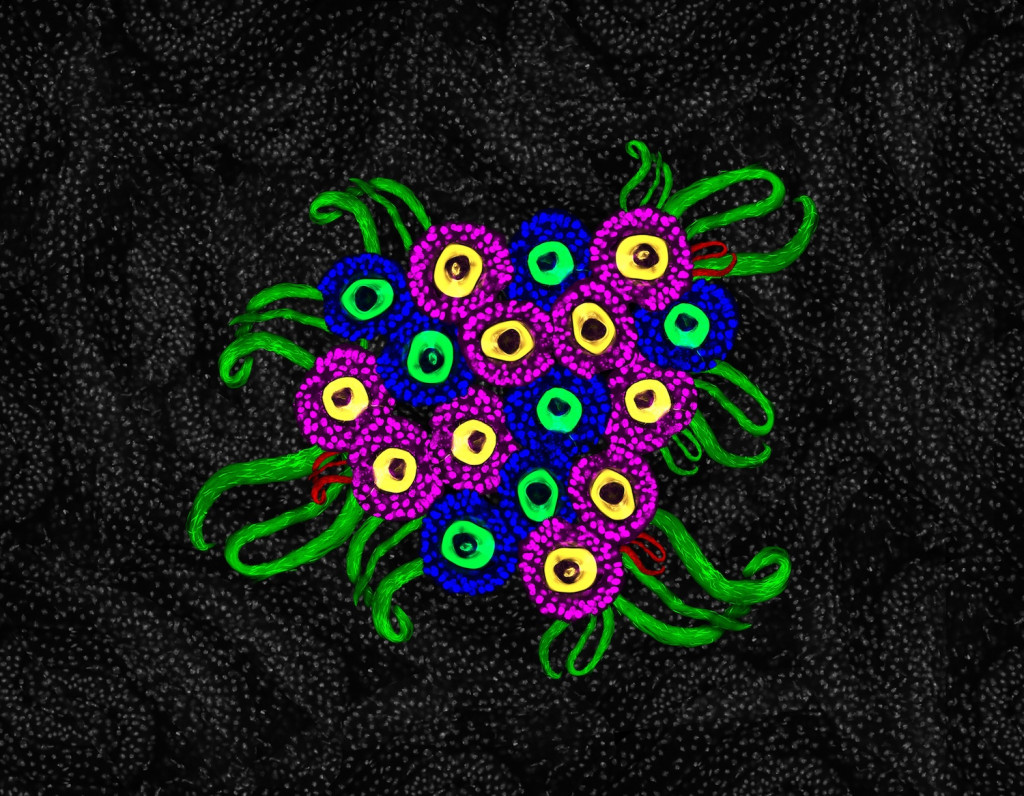
This bouquet of “flowers” is a collage of sperm storage organs (shown in green and red) from female fruit flies. Pink and blue colors mark spermatheca; green and yellow circles are moving sperm cells. The visualization helps researchers characterize the effect of warm temperatures (typically negative) on the flies’ ability to produce sperm. Ana Caroline Gandara, assistsant scientist, Genetics Confocal microscope
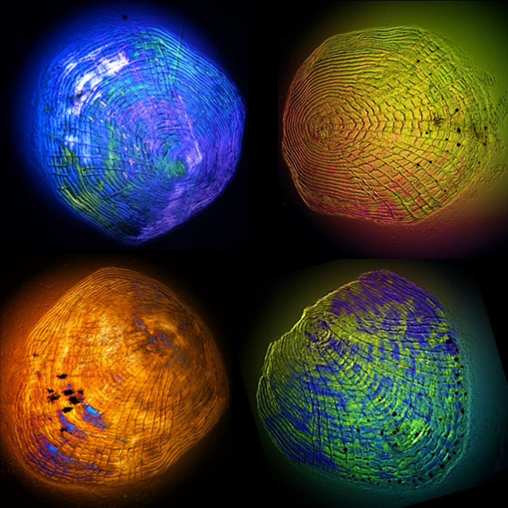
Sheets of cells migrate away from the edge of four scales taken from fathead minnows. In a dish in the lab, the cells slough off and move away as they would across the wound of a live fish. By doping the dish with different chemicals, researchers can study the way environmental contaminants affect wound-healing in the wild. Serena George, graduate student, Veterinary Medicine and Molecular and Environmental Toxicology Light microscope
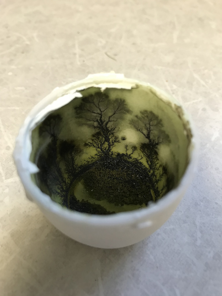
Sealed in a heated crucible for weeks in the lab of chemistry professor Robert Hamers, a tiny forest of trees grew from a glittering forest floor of individual crystals of lithium cobalt oxide, a compound used as the positive electrode in lithium-ion batteries. Robert Hamers, professor, Chemistry= Smartphone
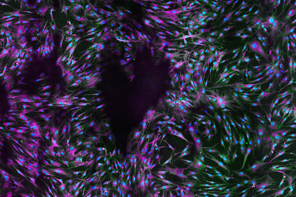
Serendipity cleared a heart shape from these human lung cells, marked by light blue nuclei. These fibroblasts — connective cells that give tissues structure — are used by researchers to study the expression of genes involved in pulmonary fibrosis, a disease that leaves severe and irreversible scarring in the lungs. Angie Tebon Oler, researcher II, Department of Medicine Confocal microscope
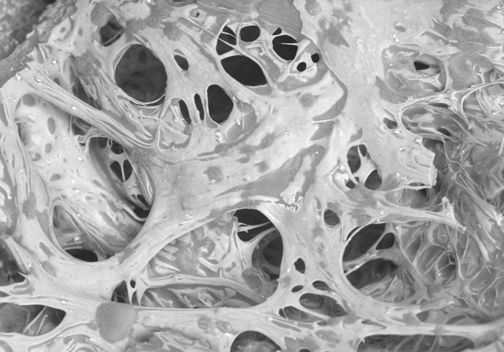
Inside your mouth isn’t the only place the magic of a cheese puff happens. The microstructures formed by proteins (lighter-colored regions) and fats (darker areas) in the interior of this cheddar puff give food science researchers clues to the development of the sensory properties of an irresistible snack. Jason M. Pronschinske, graduate student, Food Science Bil Schneider, Wilcox SEM Lab manager, Geology Rani Govindasamy-Lucey, distinguished scientist, Center for Dairy Research John J. Jaeggi, cheese industry and applications program coordinator, Center for Dairy Research Rodrigo A. Ibáñez, scientist, Center for Dairy Research Mark E. Johnson, distinguished scientist, Center for Dairy Research John A. Lucey, professor, Food Science, and director, Center for Dairy Research Scanning electron microscope
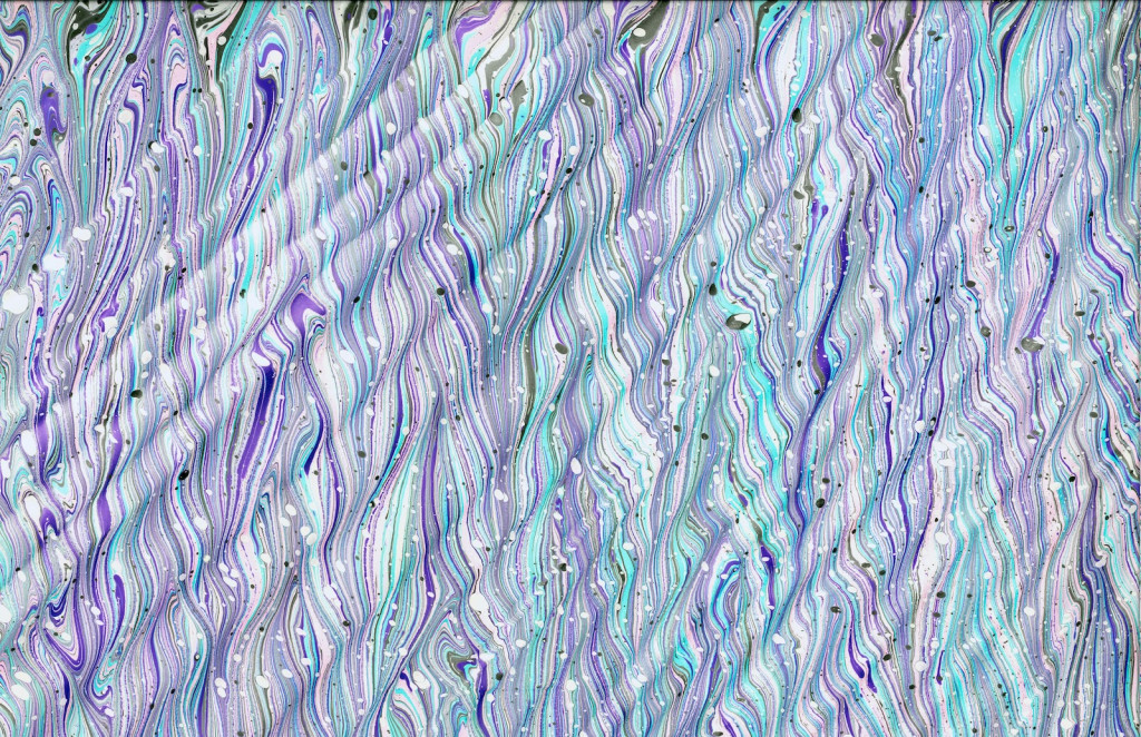
Researchers in UW–Madison’s Applied Math, Engineering and Physics Laboratory study complex interactions at the interfaces of different liquids by making marbling art, in which complex patterns in paint floated on a water bath are transferred onto paper. This scanned paper sheet — combining patterns known as “Spanish wave” and “antique” — was made in the lab in 2023 during a study of how the bath’s viscosity affects the stability of the patterns in the floating paint. Yue Sun, postdoctoral researcher, Mathematics Flatbed scanner
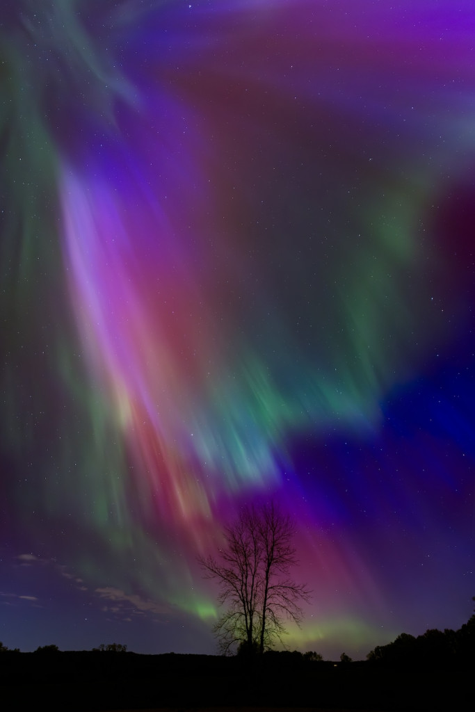
A wind of charged particles from the Sun crashed into the Earth’s magnetic field — the strongest such storm in decades — and lit up the sky over Madison on May 10 and 11, 2024. Collisions between the particles and molecules of oxygen and nitrogen produced the characteristic colors of the aurora borealis, seen here in a panorama of four combined images taken in rural Columbia County. Samuel L. Warfel, undergraduate student, Astronomy and Physics Digital camera
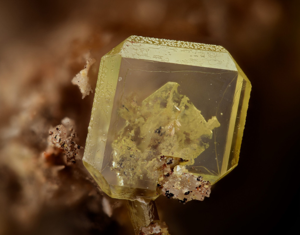
With a custom rig that moved his camera just 6 thousandths of a millimeter between photos, Prof. Joshua D. Warner took a stack of images that were combined to present the complex features of a crystal of wulfenite less than 2 millimeters wide in the otherwise impossibly sharp focus. Joshua D. Warner, assistant professor, Radiology Digital camera
The human cells in this short video, made with an epifluorescence microscope, were engineered so that the organization of particular proteins would be revealed in patterns resembling spiraling “waves” or leopard spots on the cells’ surfaces. Controlling the organization of molecules in cells can help scientists study cellular process or even find ways to prevent the sort of disorganization that can cause developmental diseases or cancer.
Scott Coyle, assistant professor, Biochemistry
Chih-Chia “Eden” Chang, graduate student, Biophysics
On average, people with Down syndrome develop Alzheimer’s disease about 30 years earlier than neurotypical people. UW–Madison researchers are tracking the progression of Alzheimer’s in Down syndrome patients by scanning their brains for the development of clumps, called plaques, of a protein called beta-amyloid. This animation made by combining the PET scans of many Down syndrome patients collected over 15 years, reveals the areas (in deepening red) in which beta-amyloid plaques developed most often.
Matthew Zammit, scientist, Waisman Center Brain Imaging Core
UW–Madison’s annual Cool Science Image Contest recognizes the technical and creative skills required to capture images, videos and other media that reveal something about the world around us while also leaving an impression with their beauty or ability to induce wonder. The contest is sponsored by Madison’s Promega Corp., with additional support from UW–Madison’s Office of Strategic Communication.
Winning entries are shared widely in UW–Madison venues, and all entries are showcased at campus science outreach events and in academic and lab facilities around campus throughout the year. See last year’s winners.
The 2024 contest judges were:
- Steve Ackerman, emeritus professor of atmospheric and oceanic sciences
- Kevin Eliceiri, professor of medical physics and director, Laboratory for Optical and Computational Instrumentation
- Michael P. King, visual communications specialist, College of Agricultural and Life Sciences
- Kara Rogers, senior editor of biomedical sciences at Encyclopaedia Britannica
- Ahna Skop, professor of genetics
- Kelly Tyrrell, assistant vice chancellor, content strategy, Office of Strategic Communication


