
‘Lazy eye’ may bully the brain into altering its wiring
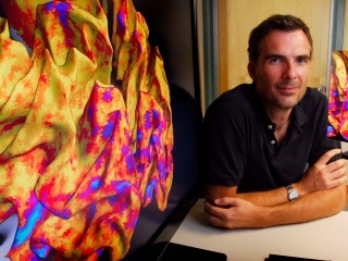
Each colored pathway represents a connection between the eyes and the visual processing areas in the brain of a person with amblyopia. Courtesy Rokers Vision Lab
Colorful and expressive, the eyes are central to the way people interact with each other, as well as take in their surroundings.
That makes amblyopia — more commonly known as “lazy eye” — all the more obvious, but the physical manifestation of the most common cause of vision problems among children the world over is actually a brain disorder.
“Most often in amblyopia patients, one eye is better at focusing,” says Bas Rokers, a University of Wisconsin–Madison psychology professor. “The brain prefers the information from that eye, and pushes down the signal coming from the other, ‘lazy’ eye. In a way, it’s better to think of the better eye as a bully, rather than the poorer eye as lazy.”
As the brain develops its preference for the dominant eye’s input, it alters its connections to the weaker eye, according to a study Rokers and colleagues published this week in a special edition of the journal Vision Research.
Bas Rokers
“If you continually have that bullying happening, that changes the signals coming from the lazy eye,” Rokers says. “We wondered, if you don’t have as many signals traveling back and forth, does that come with a physical change in those passageways?”
Using a brain scanning method called diffusion-weighted imaging, the researchers mapped three sets of pathways known to carry visual information from the eyes to the brain. In people with amblyopia, the researchers saw water diffusing more easily down the brain’s visual pathways.
“What we think may be happening in amblyopia is that the conductive sheath around neurons becomes thinner,” Rokers says. “In order to conduct information from one location to another, neurons have a sheath of material called myelin around them to insulate and speed up processing. When the myelin is thinner, there is less of it in the way and the water diffuses more easily.”
This understanding of the structural effects of amblyopia may improve treatments for amblyopia and similar vision disorders in which sufferers have trouble judging distance and location of objects in parts of their visual field.
“You don’t see any adults walking around with patched eyes, because adults’ brains are less plastic, less trainable, and we think the patch approach doesn’t have any effect late in life.”
Bas Rokers
The most common medical response to lazy eye is to correct the cause — most often muscular misalignment of the eyes, but sometimes a misshapen lens — through surgery, and put a patch over the amblyope’s strong eye to force the brain to adapt to using the formerly lazy one. But that treatment is usually limited to children.
“You don’t see any adults walking around with patched eyes, because adults’ brains are less plastic, less trainable, and we think the patch approach doesn’t have any effect late in life,” says Rokers, whose group’s work has been funded by the Wisconsin Alumni Research Foundation and the Netherlands Organization for Scientific Research. “But that belief is changing, and this diffusion-weighted imaging approach will help us understand whether, and how much, brain training treatments work.”
It will also aid in the development of new treatments — like some Rokers and ophthalmologists are developing using video games and virtual reality headsets.
“You can put patients in the scanner and see if your treatment actually has an effect,” Rokers says. “We haven’t tried many different kinds of treatments, but with a way like this to assess success, you can reward experimentation.”
-
Interwoven
How Indigenous knowledge and science can work together to communicate about climate.
-
UW–Madison’s reach throughout Wisconsin adds up to $38.9 billion a year
The university supports more than 287,000 jobs and helps drive private-sector growth.
-
‘Being part of something bigger than you’ with Badger Volunteers
UW–Madison students head into the local community to find purpose and make a difference.
-
UW–Madison’s Tech Exploration Lab: Where the classroom meets the real world
The lab is built around a simple expectation: Students come to build, test and refine projects with real problems in mind.


