New Faculty Focus: Elizabeth R. Wright
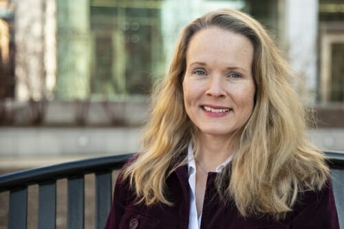
Name: Elizabeth R. Wright
Title: Professor and director of cryo-EM facility
Hometown: Pinehurst, North Carolina
Where did you grow up? My dad was a U.S. Army officer, so we lived all over the United States and across the globe. It’s hard to say where my favorite places were because there were and are so many wonderful places and people, as well as great times and memories.
Educational/professional background: B.S.in Biology and Chemistry, Columbus State University (1995 and 1997); Ph.D. in Chemistry, Emory University (2002) and postdocs at University of Southern California, Los Angeles (2003) and California Institute of Technology (2003-2008). Previously was associate professor in the Division of Infectious Disease, Department of Pediatrics, Emory University School of Medicine and Director of Emory’s Robert P. Apkarian Integrated Electron Microscopy Core.
How did you get into your field of research? I developed a love of imaging and structural biology in graduate school. I should mention, that as an undergraduate, I almost had enough credit hours in art and art history for another major! While I was a budding chemist, I did not want to lose my talent and thoughtfulness about the visual arts and I believe the craft involves producing something meaningful. Therefore, as I was considering research groups for my Ph.D., I thought about several things. What did I want to learn? Who did I want to become? I joined a group that provided me with foundations in molecular biology, protein design and engineering, and biophysical methods. Cryo-electron microscopy (cryo-EM) is a biophysical method and technology that allows us to obtain atomic, near-atomic, and macromolecular resolution images (structures) of biological molecules, like proteins and nucleic acids. The overall concept is similar to a microscope a child would use but instead of visible light passing through the specimen to form an image, it is a focused beam of electrons.
Main goal(s) of your current research program: My research interests are varied but all revolve around the development and use of advanced light and electron microscopy imaging technologies to study complex biology. I am intrigued by how mammalian cells, bacteria, and viruses assemble and function. Using cryo-EM, we can collect time-resolved ‘snap shots’ of biological systems to follow structural changes that occur during both normal functional and diseased states. This structural insight can provide us with a wealth of information that can be translated to the development of drug targets, therapeutics, and treatments.
What attracted you to UW–Madison? I came to UW–Madison to join the faculty in the Department of Biochemistry and to direct the department’s new cryo-EM facility that will serve as a resource for all of campus. The Department of Biochemistry’s vision for this facility really drew me to UW–Madison. We are not just thinking about the present state of structural biology and the field of cryo-EM, but about making investments that will shape the next several decades of research in the fields of structural biology, biochemistry, cell biology, and medicine and build a community of investigators across the UW–Madison campus.
What was your first visit to campus like? During my first visit I found the campus to be extremely vibrant, with students bustling around to classes, labs, and study spaces. I really enjoyed meeting with people in the department, including students, postdocs, staff, and the faculty. My second visit provided me with a taste of a Wisconsin winter. When I arrived around noon it was a pleasant 50ºF, but by 7 p.m. it was 15º and quite windy! Coming from Atlanta, I was frostbitten. That said, it was the first significant snowstorm of the winter and it was nice to see everyone excited about the snow. It was also pretty incredible to see the lakes completely frozen over! Who knew that frozen custard and ice cream are staples in midwinter!
Favorite place on campus? I love nature and the outdoors so being close to the lake for walks and reflection is perfect. My family and I enjoy regular visits to the Microbial Sciences building to check on the ant colony. Of course, the Babcock Dairy Store is ever popular.
What are you most enjoying so far about working here? I have enjoyed meeting with and working with many people across the UW–Madison campus. There is ferocious loyalty to UW–Madison among members of the community, which really brings people together to support cross-cutting initiatives. On Wisconsin! Go Badgers!
Do you feel your work relates in any way to the Wisconsin Idea? If so, please describe how. I believe a lot of the work in my lab relates to the Wisconsin Idea. In the laboratory and in collaboration with other research groups, we develop technologies for imaging, sample handling and manipulation, and data analysis. Our efforts readily feed into technology transfer because other researchers will benefit from the advances we make to hardware, software, and workflows. On the biological side of the laboratory, structures we solve by cryo-EM support our capacity to develop new therapeutics, vaccines, and antimicrobials that could have a global impact.
I also have a passion for science outreach with children of all ages. Simple microscopy lessons lend themselves to providing students with opportunities to gain a greater appreciation of the world around them. One of the most enjoyable classroom labs is to have students use their eyes, a magnifying glass, a light microscope, and then an electron microscope to view (or image) the same object at different scales and resolution levels. It is with these snapshots that we may instill in children a sense of awe, wonder, and curiosity about the complexity of life and all living things.
What’s something interesting about your area of expertise you can share that will make us sound smarter at parties? For cryo-EM, we freeze, or vitrify, the aqueous samples at approximately -183° Celsius. The rapid freezing process supports the formation of a non-crystalline phase of ice that is amorphous, vitreous (glass-like) ice. When the electron beam from the microscope passes through the liquid nitrogen-cooled sample, the ice and the specimen move like a fluid without breaking. We all learn early in school about the three phases of water — solid (ice), liquid (water), gas (steam) — but, there are actually multiple phases that ice alone transitions through! Super cool science!
Hobbies/other interests: I am also an artist and enjoy painting, landscapes in particular, and photography. Even though I chose science as a career, working in the field of imaging science allows me to retain my artistic eye.


