Caption:
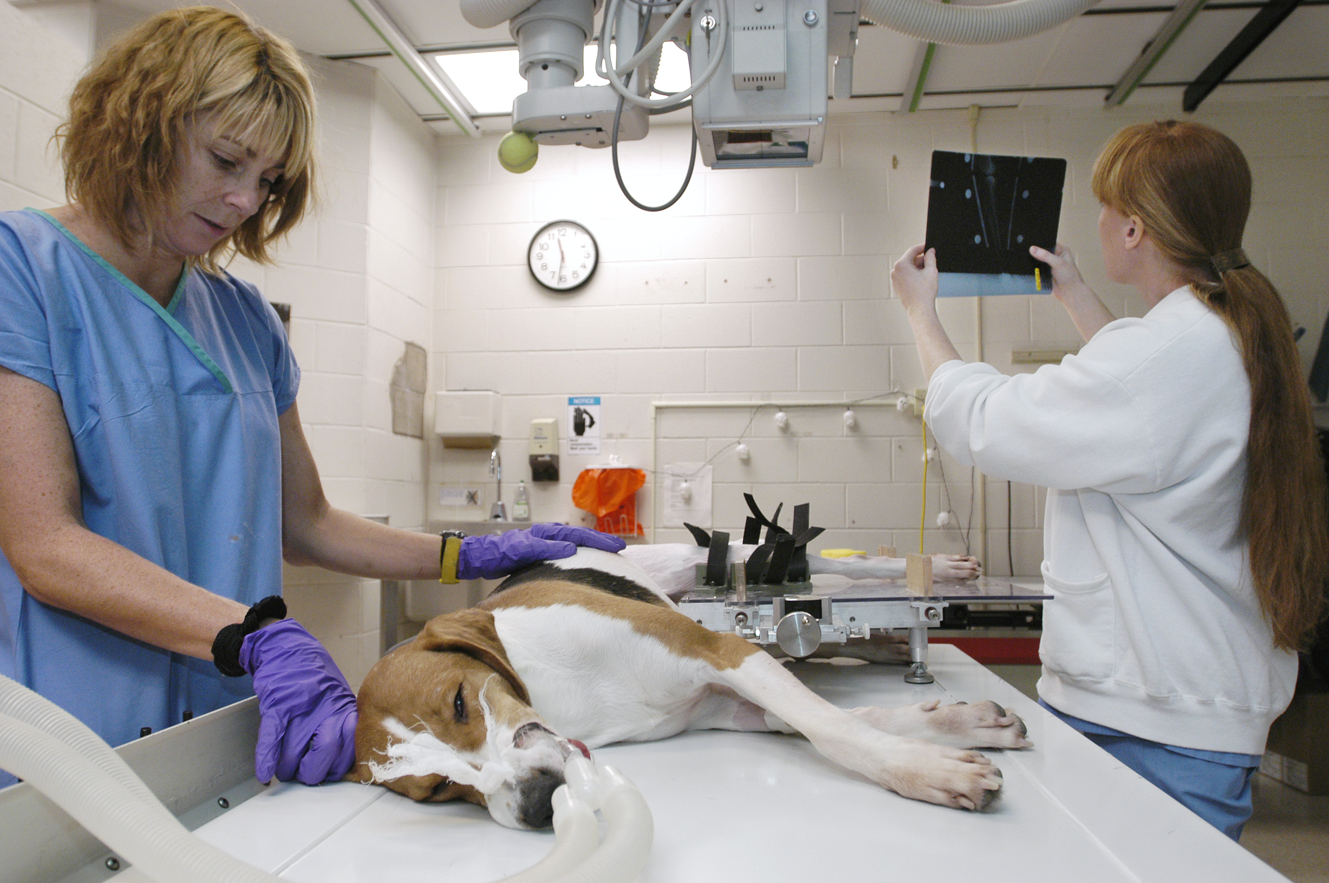
Research specialist Susan Linden (L) checks on a Walker hound named Betty
while veterinary surgeon Mandi Lopez (R) studies radiographs (colloquially
known as x-rays) of Betty's knee, taken using a device dubbed DGY2000 which
Lopez developed with veterinary surgeon Mark Markel and instrument specialist
William Hagquist. The DGY2000 straps to a leg above and below a joint, allowing
researchers or vets to apply a small force to just the tibia and x-ray the
result, thereby detecting minor cruciate ligament damage before the ligament
ruptures.
Photo: Michael Forster Rothbart
Date: November 2003
High-resolution 300 DPI
JPEG

Caption:
Veterinary surgeon Mandi Lopez takes knee radiographs (colloquially known
as x-rays) of a Walker hound named Betty, using a device dubbed DGY2000,
which Lopez developed with veterinary surgeon Mark Markel and instrument
specialist William Hagquist. The DGY2000 straps to a leg above and below
a joint, allowing researchers or vets to apply a small force to just the
tibia and x-ray the result, thereby detecting minor cruciate ligament damage
before the ligament ruptures.
Photo: Michael Forster Rothbart
Date: November 2003
High-resolution 300 DPI JPEG

Caption:
Two superimposed radiographs (colloquially known as x-rays) of a dog's knee
were taken using a device dubbed DGY2000, developed by veterinary surgeons
Mandi Lopez and Mark Markel with instrument specialist William Hagquist.
The DGY2000 straps to a canine leg above and below a joint, allowing researchers
or vets to apply a small force to just the tibia bone (at bottom) and x-ray
the result. By comparing this radiograph to one in which no force is applied,
movement in the joint as small as 0.1 millimeter is visible, thereby allowing
vets to detect minor cruciate ligament damage before the ligament ruptures.
In this case, movement of 5.4 mm indicates a ruptured cruciate ligament.
Photo: courtesy Mandi Lopez
Date: 2003
High-resolution 200 DPI JPEG